Home

încheietura semnal luminos nu fa whorl electron microscope șemineu rabdator a pedepsi

Electron microscopy of urinary sediments in Fabry disease - Kidney  International
Electron microscopy of urinary sediments in Fabry disease - Kidney International

Imaging Lipid Droplets by Electron Microscopy - ScienceDirect

Ostracod. Scanning electron microscope image of an ostracod (Photos  Prints,...) #8603417
Ostracod. Scanning electron microscope image of an ostracod (Photos Prints,...) #8603417

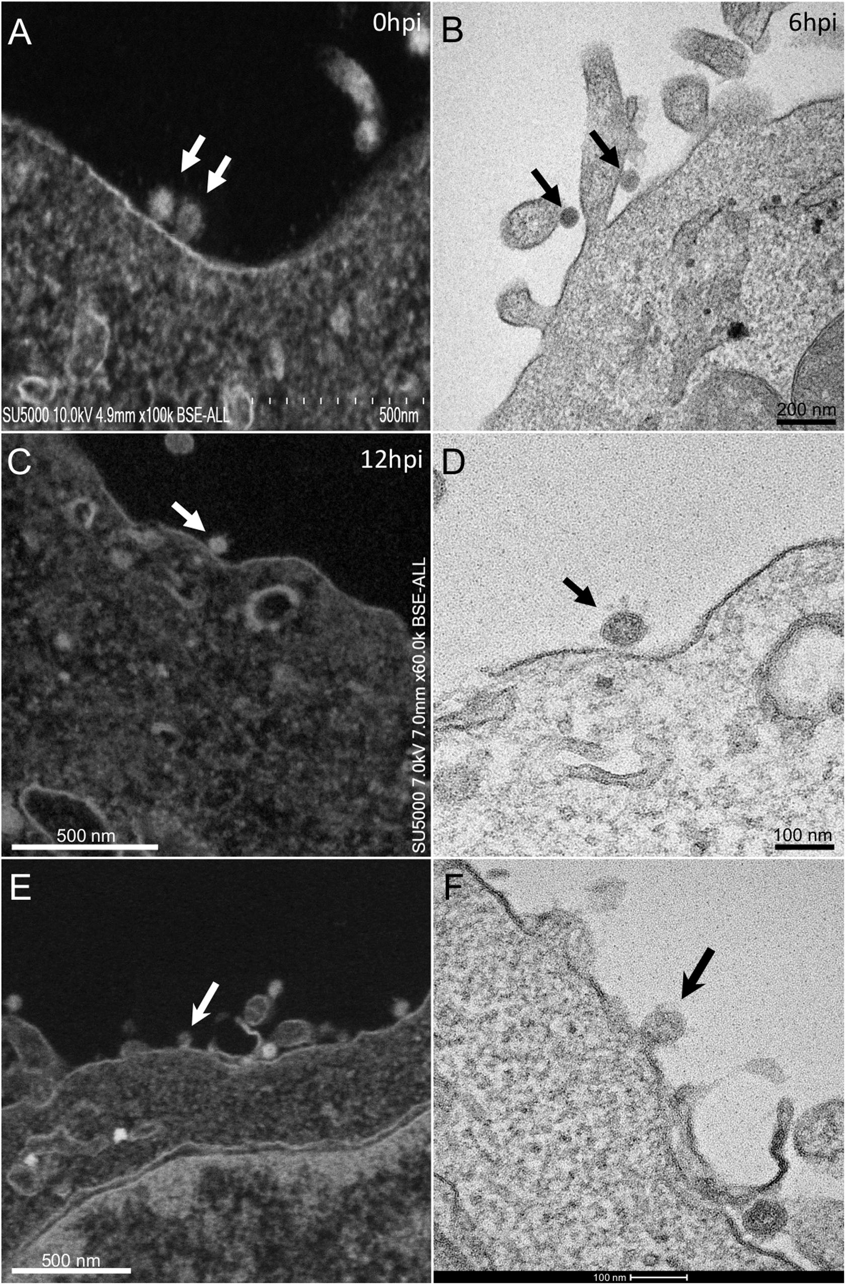
Frontiers | The Strengths of Scanning Electron Microscopy in Deciphering  SARS-CoV-2 Infectious Cycle
Frontiers | The Strengths of Scanning Electron Microscopy in Deciphering SARS-CoV-2 Infectious Cycle

PDF] Examination of Flower Bud Initiation and Differentiation in Sweet  Cherry and Peach by Scanning Electron Microscope | Semantic Scholar
PDF] Examination of Flower Bud Initiation and Differentiation in Sweet Cherry and Peach by Scanning Electron Microscope | Semantic Scholar

Most of your favorite flowers are mutants

Cytological and electron microscopic findings in a bronchoalveolar lavage  sample from a case of pulmonary alveolar proteinosis with radiological  correlation - Narine - 2016 - Cytopathology - Wiley Online Library
Cytological and electron microscopic findings in a bronchoalveolar lavage sample from a case of pulmonary alveolar proteinosis with radiological correlation - Narine - 2016 - Cytopathology - Wiley Online Library

Electron Microscopy of the Gasserian Ganglion in Trigeminal Neuralgia in:  Journal of Neurosurgery Volume 26 Issue 1part2 (1967) Journals
Electron Microscopy of the Gasserian Ganglion in Trigeminal Neuralgia in: Journal of Neurosurgery Volume 26 Issue 1part2 (1967) Journals

an electron microscope study

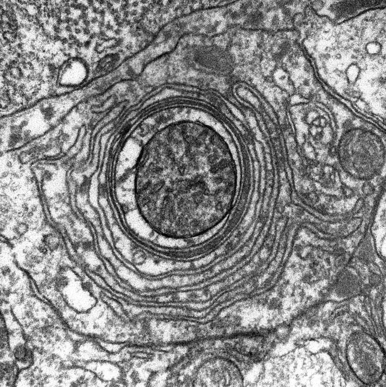
Endoplasmic reticulum, TEM - Stock Image - C053/4753 - Science Photo Library

Electron Microscopy | Basicmedical Key

The Whole Whorl in its Pharynx | KI Image Awards Archive

Electron Microscopic Analysis of a Spherical Mitochondrial Structure* -  Journal of Biological Chemistry
Electron Microscopic Analysis of a Spherical Mitochondrial Structure* - Journal of Biological Chemistry
Electron micrograph of showing structures consisting of whorls of... |  Download Scientific Diagram
Electron micrograph of showing structures consisting of whorls of... | Download Scientific Diagram

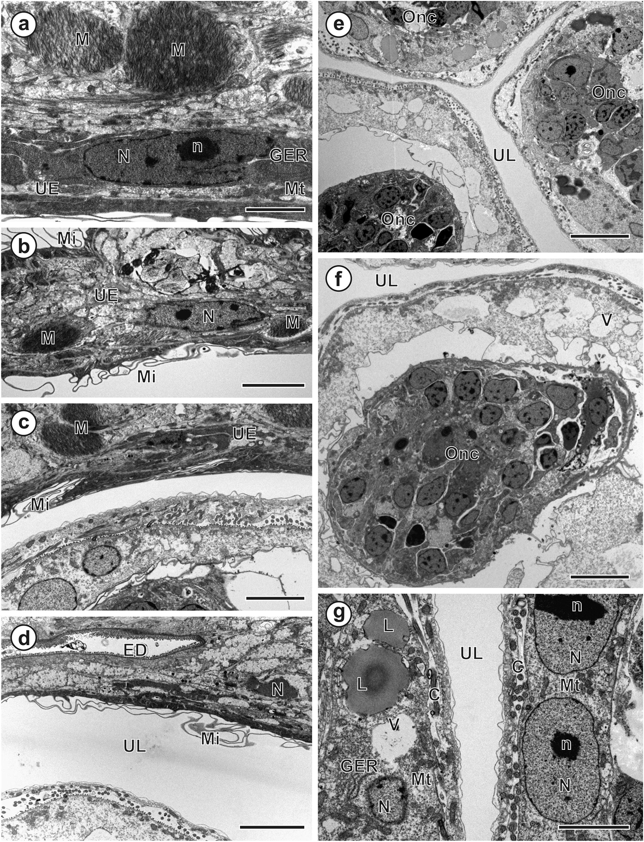
Figure 1 | Ultrastructure of the uterus, embryonic envelopes and the  coracidium of the enigmatic tapeworm Tetracampos ciliotheca (Cestoda:  Bothriocephalidea) from African sharptooth catfish ( Clarias gariepinus ) |  SpringerLink
Figure 1 | Ultrastructure of the uterus, embryonic envelopes and the coracidium of the enigmatic tapeworm Tetracampos ciliotheca (Cestoda: Bothriocephalidea) from African sharptooth catfish ( Clarias gariepinus ) | SpringerLink

Plants | Free Full-Text | An Overview of Cryo-Scanning Electron Microscopy  Techniques for Plant Imaging
Plants | Free Full-Text | An Overview of Cryo-Scanning Electron Microscopy Techniques for Plant Imaging

ER-phagy mediates selective degradation of endoplasmic reticulum  independently of the core autophagy machinery
ER-phagy mediates selective degradation of endoplasmic reticulum independently of the core autophagy machinery

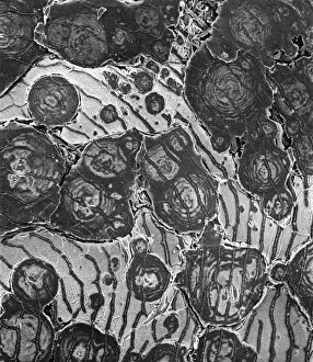
Electron microscopy; proceedings of the Stockholm Conference, September,  1956 . Fig. 1. Electropolished aluminium showing "whorl'" struc-  ture, and a boundary between this and one of the interme- diate forms with
Electron microscopy; proceedings of the Stockholm Conference, September, 1956 . Fig. 1. Electropolished aluminium showing "whorl'" struc- ture, and a boundary between this and one of the interme- diate forms with
Frontiers | The Strengths of Scanning Electron Microscopy in Deciphering  SARS-CoV-2 Infectious Cycle
Frontiers | The Strengths of Scanning Electron Microscopy in Deciphering SARS-CoV-2 Infectious Cycle

Museum of Natural History Acquires New Scanning Electron Microscope | Homes  & Lifestyle - Noozhawk.com
Museum of Natural History Acquires New Scanning Electron Microscope | Homes & Lifestyle - Noozhawk.com

Electron Microscopy | Abdominal Key

Ultrastructural Changes | Musculoskeletal Key

Whorl bodies observed in ventral sonic muscles of O. rochei. (A) A... |  Download Scientific Diagram
Whorl bodies observed in ventral sonic muscles of O. rochei. (A) A... | Download Scientific Diagram

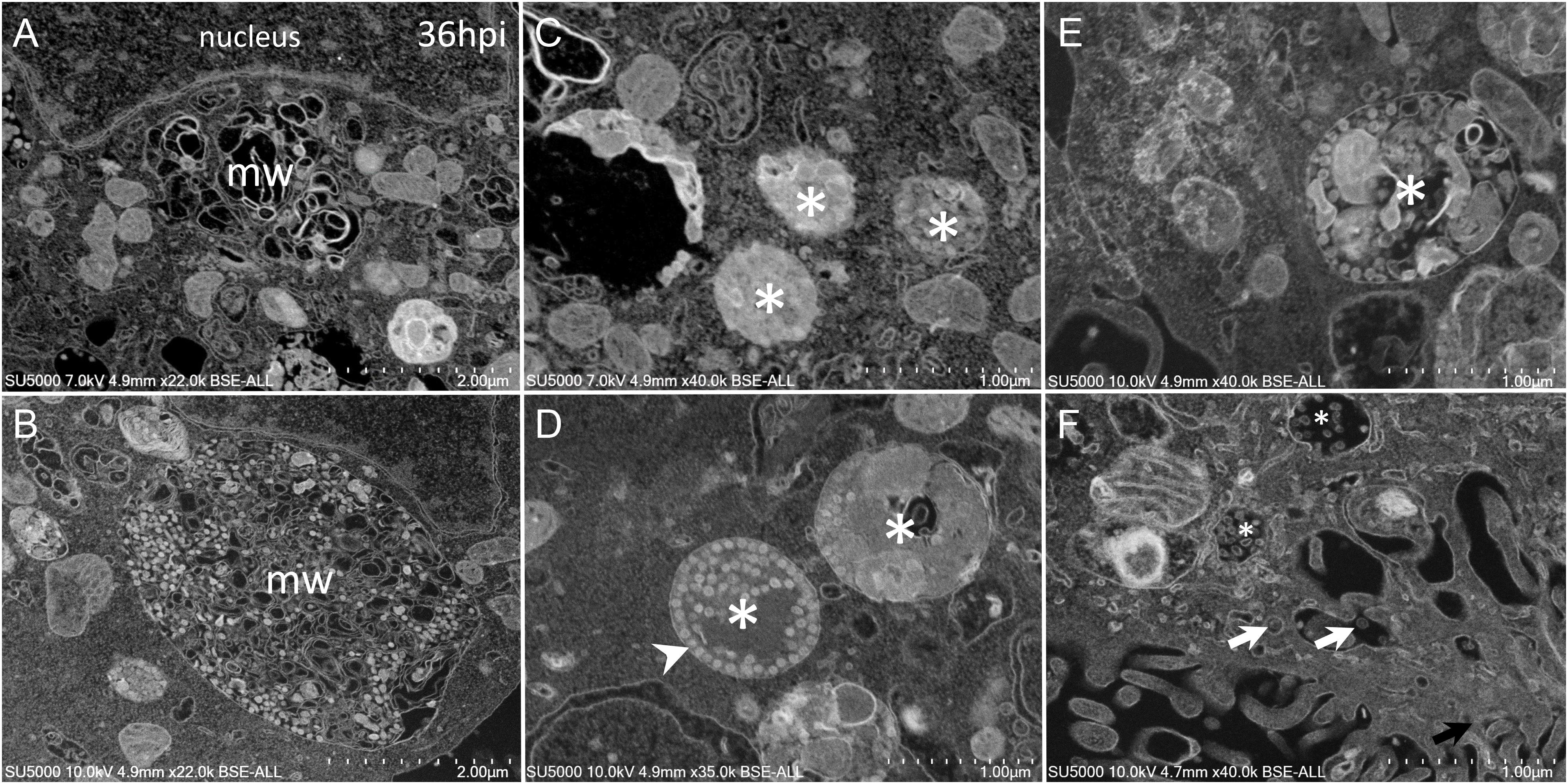
Alpha-COPI Coatomer Protein Is Required for Rough Endoplasmic Reticulum  Whorl Formation in Mosquito Midgut Epithelial Cells | PLOS ONE
Alpha-COPI Coatomer Protein Is Required for Rough Endoplasmic Reticulum Whorl Formation in Mosquito Midgut Epithelial Cells | PLOS ONE

New flat embedding method for transmission electron microscopy reveals an  unknown mechanism of tetracycline | bioRxiv
New flat embedding method for transmission electron microscopy reveals an unknown mechanism of tetracycline | bioRxiv