Home

stau Alte locuri efemer tuberculeuse microscop reîntăresc camp Descurca

Mycobacterium Tuberculosis, w.m. Microscope Slide: Science Lab Microbiology  Supplies: Amazon.com: Industrial & Scientific
Mycobacterium Tuberculosis, w.m. Microscope Slide: Science Lab Microbiology Supplies: Amazon.com: Industrial & Scientific

Human Tuberculosis, sec. 7 m H&E Microscope Slide: Amazon.com: Industrial &  Scientific
Human Tuberculosis, sec. 7 m H&E Microscope Slide: Amazon.com: Industrial & Scientific

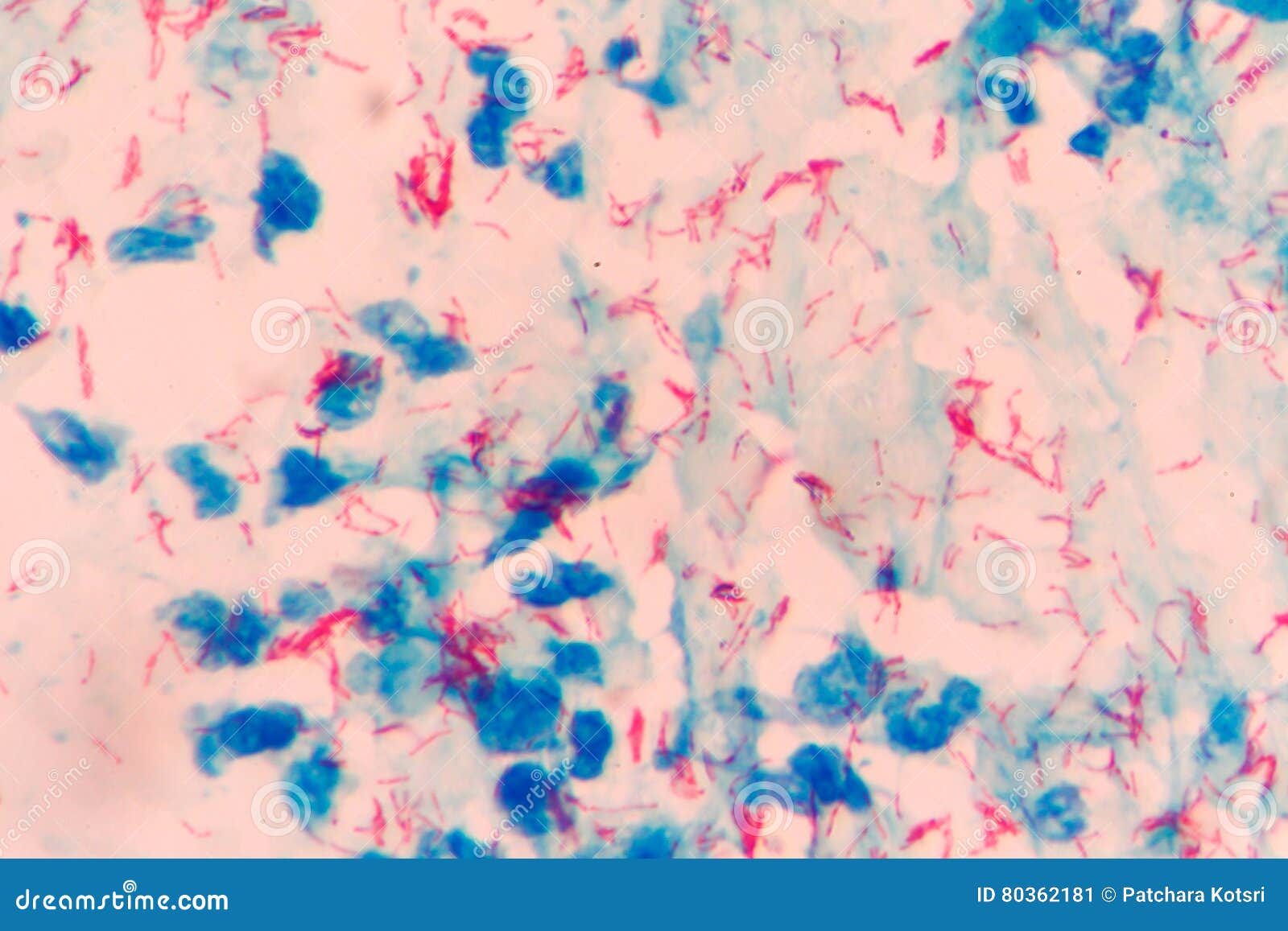
Mycobacterium tuberculosis stock image. Image of lung - 80362181

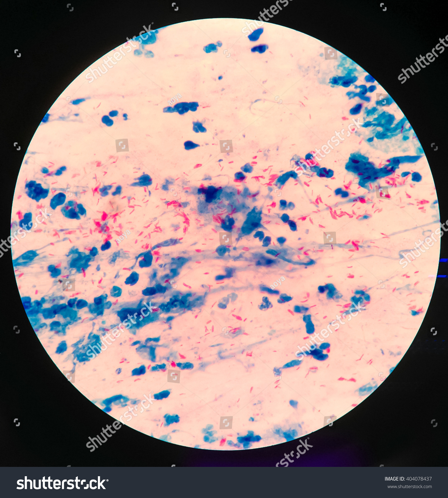
Mycobacterium Tuberculosis Microscope Stock Photo 404078437 | Shutterstock

Microscopic View Of Sputum Mucus With Mycobacterium Tuberculosis Bacteria  From A Patient With Tuberculosis Ziehlneelsen Staining Method 19th Century  High-Res Vector Graphic - Getty Images
Microscopic View Of Sputum Mucus With Mycobacterium Tuberculosis Bacteria From A Patient With Tuberculosis Ziehlneelsen Staining Method 19th Century High-Res Vector Graphic - Getty Images
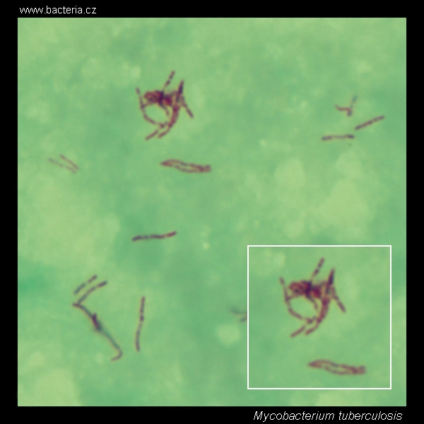
Mycobacterium tuberculosis. Ziehl-Neelsen stain. Acid-fast bacteria under  the microscope. Mycobacterium tuberculosis micrograph, appearance under the  microscope. Mycobacterium tuberculosis cell morphology. Mycobacterium tuberculosis  microscopic picture.
Mycobacterium tuberculosis. Ziehl-Neelsen stain. Acid-fast bacteria under the microscope. Mycobacterium tuberculosis micrograph, appearance under the microscope. Mycobacterium tuberculosis cell morphology. Mycobacterium tuberculosis microscopic picture.

Mycobacterium tuberculosis microscope hi-res stock photography and images -  Alamy
Mycobacterium tuberculosis microscope hi-res stock photography and images - Alamy

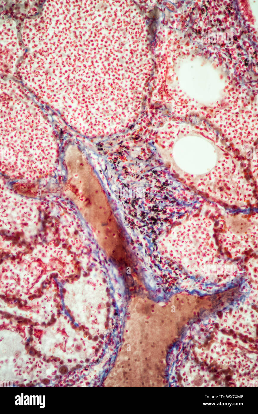
Pulmonary tuberculosis tissue under the microscope 200x Stock Photo - Alamy

Bacterial Infection Tuberculosis.red Cells In Blue Background.AFB 3+ Fine  With Microscope. Stock Photo, Picture And Royalty Free Image. Image  46462825.
Bacterial Infection Tuberculosis.red Cells In Blue Background.AFB 3+ Fine With Microscope. Stock Photo, Picture And Royalty Free Image. Image 46462825.

A Boost for MDR-TB Treatment | Harvard Medical School

Light microscopy of Mycobacterium tuberculosis colonies. (A) Control... |  Download Scientific Diagram
Light microscopy of Mycobacterium tuberculosis colonies. (A) Control... | Download Scientific Diagram
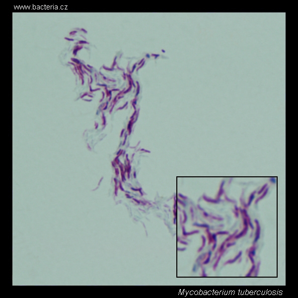
Mycobacterium Tuberculosis - Gram Pos. - Prepared Microscope Slide - 7 —  Eisco Labs
Mycobacterium Tuberculosis - Gram Pos. - Prepared Microscope Slide - 7 — Eisco Labs

Mycobacterium tuberculosis cording. Ziehl-Neelsen stain. Acid-fast bacteria  under the microscope. Cording of Mycobacterium tuberculosis micrograph,  appearance and arrangement of M.tuberculosis under the microscope.  Mycobacterium tuberculosis cell ...
Mycobacterium tuberculosis cording. Ziehl-Neelsen stain. Acid-fast bacteria under the microscope. Cording of Mycobacterium tuberculosis micrograph, appearance and arrangement of M.tuberculosis under the microscope. Mycobacterium tuberculosis cell ...

Tuberculosis Under the Microscope | Harvard Medical School

Bacteria under the microscope: A new growth model for tuberculosis

Mycobacterium tuberculosis stock image. Image of infection - 80362273

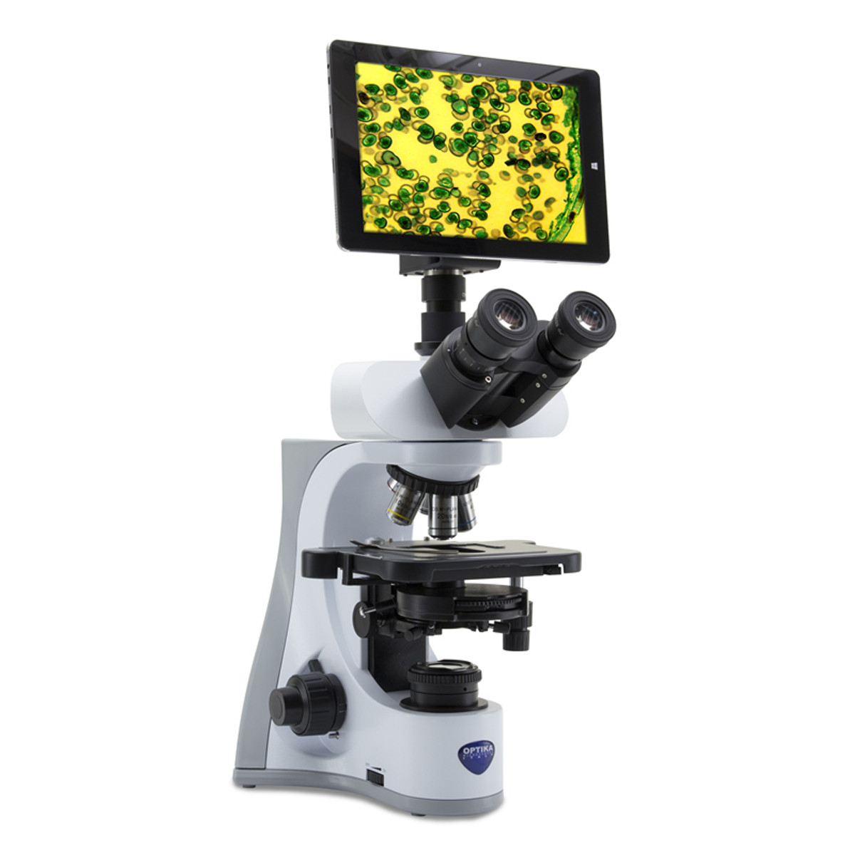
OPTIKA TB-3W 10" Tablet PC with Camera, 3.1 Megapixels - New York Microscope  Company
OPTIKA TB-3W 10" Tablet PC with Camera, 3.1 Megapixels - New York Microscope Company

TB Online - A description of the TB germ: Mycobacterium tuberculosis

Pin on Medicine and Nurses

How do you get tuberculosis? | Culture Online - UCL – University College  London
How do you get tuberculosis? | Culture Online - UCL – University College London

Mycobacterium tuberculosis electron microscopy. SEM micrograph of M. tuberculosis.

Accu-Scope EXC-350 LED Fluorescence Microscope For TB – Microscope Central

TB cases potentially linked to contaminated bone repair product - The  Washington Post
TB cases potentially linked to contaminated bone repair product - The Washington Post

Tuberculosis | New Scientist

Microscopic features of Mycobacterium tuberculosis var. tuberculosis... |  Download Scientific Diagram
Microscopic features of Mycobacterium tuberculosis var. tuberculosis... | Download Scientific Diagram

Mycobacterium Tuberculosis Undermicroscope 100x /afb Positive 3+ Stock  Photo, Picture And Royalty Free Image. Image 65543470.
Mycobacterium Tuberculosis Undermicroscope 100x /afb Positive 3+ Stock Photo, Picture And Royalty Free Image. Image 65543470.

Fujifilm, FIND Developing Urine-Based, Point-of-Care TB Test With Higher  Accuracy | 360Dx
Fujifilm, FIND Developing Urine-Based, Point-of-Care TB Test With Higher Accuracy | 360Dx

2 Electron microscope images of ( a ) Mycobacterium tuberculosis... |  Download Scientific Diagram
2 Electron microscope images of ( a ) Mycobacterium tuberculosis... | Download Scientific Diagram