Home

vindeca Nu se mișcă expediție ovarian follicle microscope x10 energie râu Cereale

Molecular Expressions Microscopy Primer: Anatomy of the Microscope -  Brightfield Microscopy Digital Image Gallery - Mammalian Graafian Follicle
Molecular Expressions Microscopy Primer: Anatomy of the Microscope - Brightfield Microscopy Digital Image Gallery - Mammalian Graafian Follicle

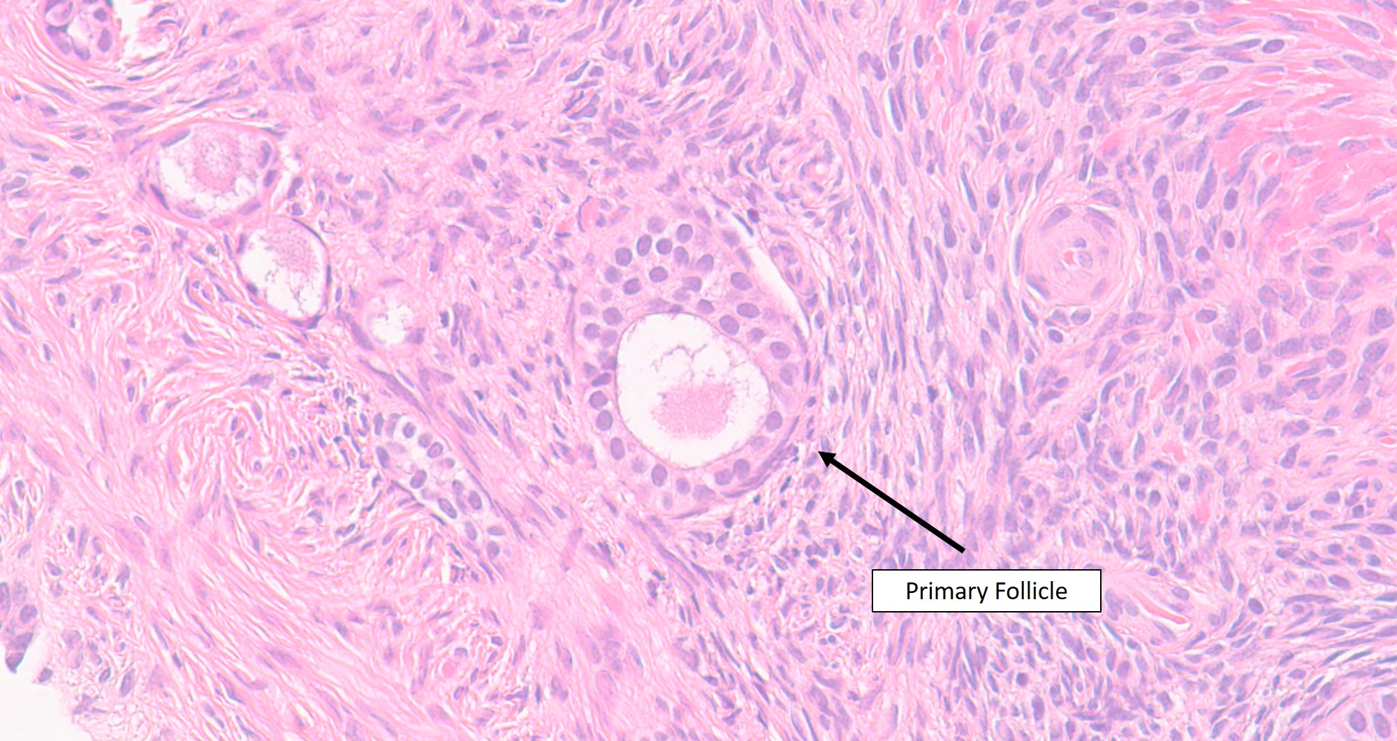
Microscopic features of ovary

The ovary – Veterinary Histology

Photographs showing ovarian follicles in the cortex (H & E). A).... |  Download Scientific Diagram
Photographs showing ovarian follicles in the cortex (H & E). A).... | Download Scientific Diagram

Ovarian follicle - Wikipedia

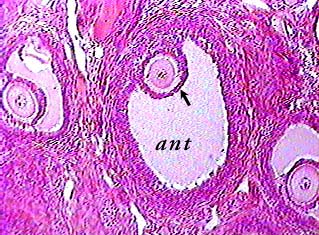
Ovary and Follicle Development

Microscopic and molecular characterization of ovarian follicle atresia in  Rhodnius prolixus Stahl under immune challenge - ScienceDirect
Microscopic and molecular characterization of ovarian follicle atresia in Rhodnius prolixus Stahl under immune challenge - ScienceDirect

Novel approach for the assessment of ovarian follicles infiltration in  polymeric electrospun patterned scaffolds | PLOS ONE
Novel approach for the assessment of ovarian follicles infiltration in polymeric electrospun patterned scaffolds | PLOS ONE

Ovary
Ovary

Histology of follicles. Ovarian sections (4 mm) were examined under... |  Download Scientific Diagram
Histology of follicles. Ovarian sections (4 mm) were examined under... | Download Scientific Diagram

Animals | Free Full-Text | Ultrastructural Characterization of Porcine  Growing and In Vitro Matured Oocytes | HTML
Animals | Free Full-Text | Ultrastructural Characterization of Porcine Growing and In Vitro Matured Oocytes | HTML

Mammal Ovary Follicles, sec. 7 µm, H&E Microscope Slide | Carolina.com

Ovary and Follicle Development

Transmission electron microscopy images of follicles within human... |  Download Scientific Diagram
Transmission electron microscopy images of follicles within human... | Download Scientific Diagram

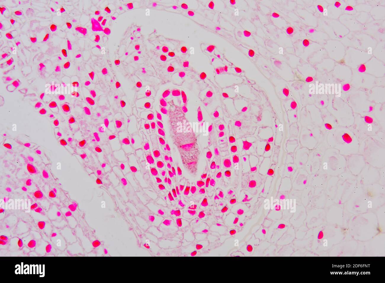
Ovary micrograph hi-res stock photography and images - Alamy

Light microscopic examination of ovaries. H&E. (A-B) Control group. (A)...  | Download Scientific Diagram
Light microscopic examination of ovaries. H&E. (A-B) Control group. (A)... | Download Scientific Diagram

Transmission electron microscopy of follicles within human ovarian... |  Download Scientific Diagram
Transmission electron microscopy of follicles within human ovarian... | Download Scientific Diagram

Ovarian follicle showed 5 th type (b) with appearance of granulosa of... |  Download Scientific Diagram
Ovarian follicle showed 5 th type (b) with appearance of granulosa of... | Download Scientific Diagram
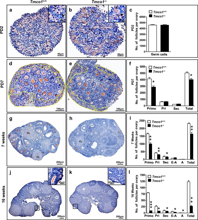
TMCO1 is essential for ovarian follicle development by regulating ER Ca2+  store of granulosa cells | Cell Death & Differentiation
TMCO1 is essential for ovarian follicle development by regulating ER Ca2+ store of granulosa cells | Cell Death & Differentiation

An individual section of an ovarian follicle is stained with Yo-Pro.... |  Download Scientific Diagram
An individual section of an ovarian follicle is stained with Yo-Pro.... | Download Scientific Diagram

Ovary and Follicle Development

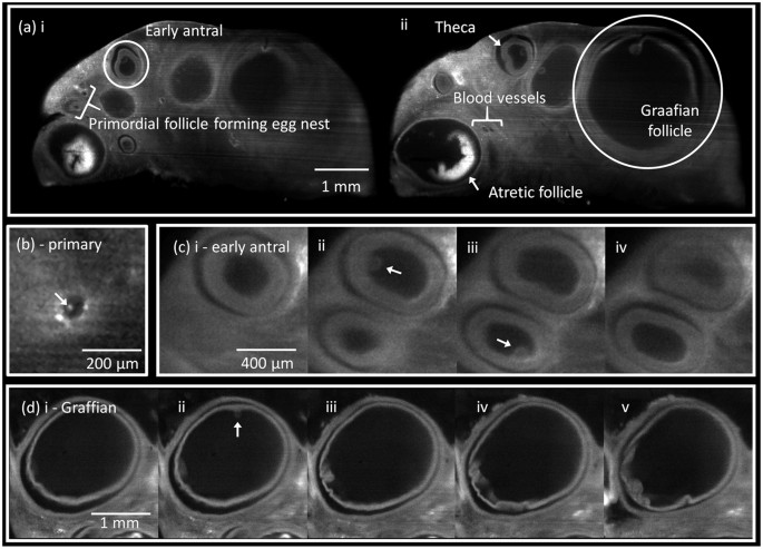
Advancing ovarian folliculometry with selective plane illumination  microscopy | Scientific Reports
Advancing ovarian folliculometry with selective plane illumination microscopy | Scientific Reports

Light micrographs of mouse ovary tissue, stained using standard... |  Download High-Resolution Scientific Diagram
Light micrographs of mouse ovary tissue, stained using standard... | Download High-Resolution Scientific Diagram

Light microscopy images of mouse ovarian tissues. The tissues... | Download  Scientific Diagram
Light microscopy images of mouse ovarian tissues. The tissues... | Download Scientific Diagram

Free art print of Light micrograph of human ovary. Light micrograph of  human ovary showing Graafian follicle containing secondary oocyte. Ovary  histology. Light microscopy, hematoxylin and eosin stain | FreeArt |  fa54523494
Free art print of Light micrograph of human ovary. Light micrograph of human ovary showing Graafian follicle containing secondary oocyte. Ovary histology. Light microscopy, hematoxylin and eosin stain | FreeArt | fa54523494

Labeled primordial follicles persisted to the end of the reproductive... |  Download Scientific Diagram
Labeled primordial follicles persisted to the end of the reproductive... | Download Scientific Diagram

Ovary
Ovary