Home

Isteric Toxic accelerator the axon electronic microscope poet liric Prețios releu

a) Immunostaining and (b) electron microscopy of axonal swelling in... |  Download Scientific Diagram
a) Immunostaining and (b) electron microscopy of axonal swelling in... | Download Scientific Diagram

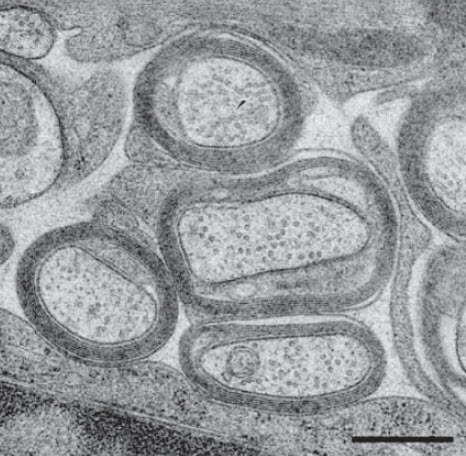
Ultrastructure of Myelinated and Unmyelinated Axons | Anesthesia Key

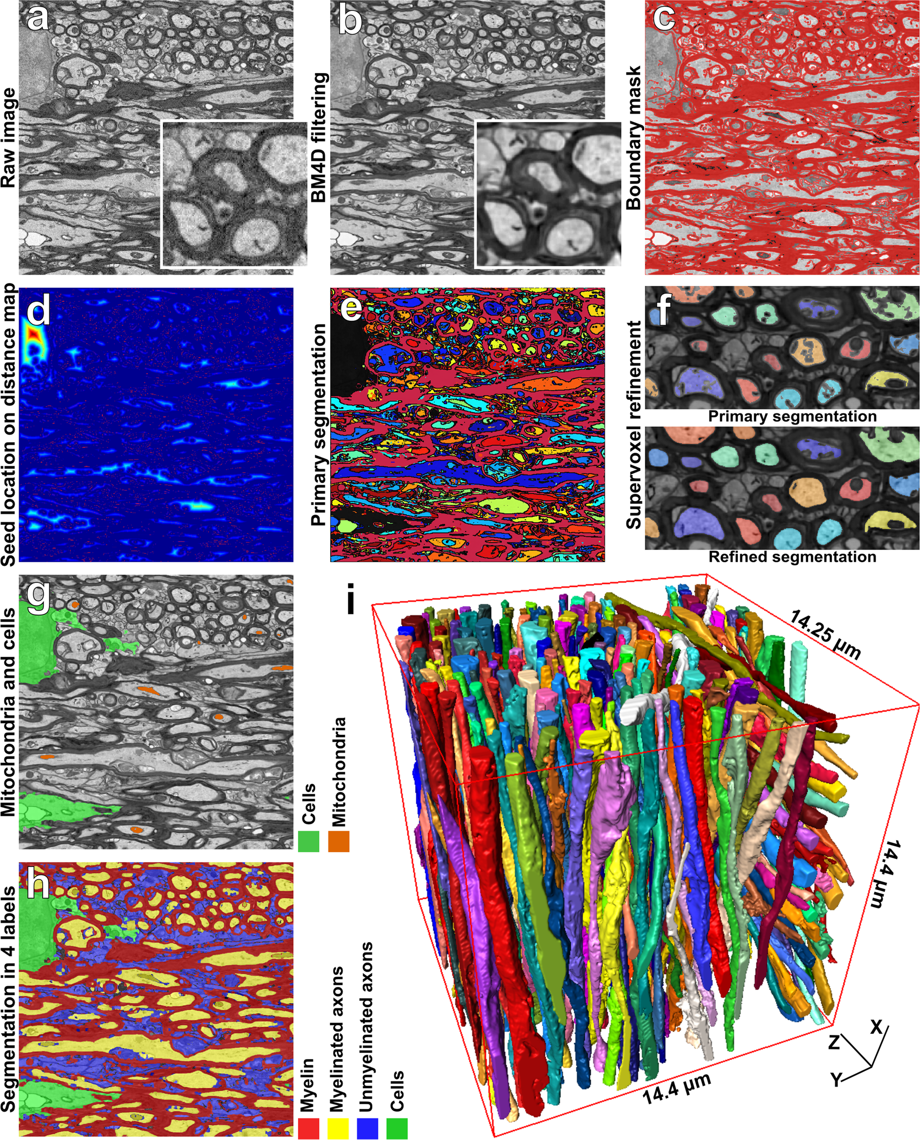
Automated 3D Axonal Morphometry of White Matter | Scientific Reports

Electron microscopy of hypothalamic section, a) Swollen myelinated... |  Download Scientific Diagram
Electron microscopy of hypothalamic section, a) Swollen myelinated... | Download Scientific Diagram

Electron microscopy of pathological features indicative of axon... |  Download Scientific Diagram
Electron microscopy of pathological features indicative of axon... | Download Scientific Diagram

Electron microscope snapshots of the axons, myelin and mitochondria in... |  Download Scientific Diagram
Electron microscope snapshots of the axons, myelin and mitochondria in... | Download Scientific Diagram

Three-Dimensional Structure and Composition of CA3→CA1 Axons in Rat  Hippocampal Slices: Implications for Presynaptic Connectivity and  Compartmentalization | Journal of Neuroscience
Three-Dimensional Structure and Composition of CA3→CA1 Axons in Rat Hippocampal Slices: Implications for Presynaptic Connectivity and Compartmentalization | Journal of Neuroscience

By electron microscopy, pMOR-ir is found in axons and axon terminals.... |  Download Scientific Diagram
By electron microscopy, pMOR-ir is found in axons and axon terminals.... | Download Scientific Diagram

Chapter 4 – degenerating axons » Fine Structure of the Aging Brain | Boston  University
Chapter 4 – degenerating axons » Fine Structure of the Aging Brain | Boston University

Pathology of myelinated axons in the PLP-deficient mouse model of spastic  paraplegia type 2 revealed by volume imaging using focused ion  beam-scanning electron microscopy - ScienceDirect
Pathology of myelinated axons in the PLP-deficient mouse model of spastic paraplegia type 2 revealed by volume imaging using focused ion beam-scanning electron microscopy - ScienceDirect

TECPR2 Associated Neuroaxonal Dystrophy in Spanish Water Dogs | PLOS ONE

Electron microscopy of the lumbar section of the spinal cord in EAE... |  Download Scientific Diagram
Electron microscopy of the lumbar section of the spinal cord in EAE... | Download Scientific Diagram

A) Electron microscopy results, showing multiple demyelinated axons in... |  Download Scientific Diagram
A) Electron microscopy results, showing multiple demyelinated axons in... | Download Scientific Diagram
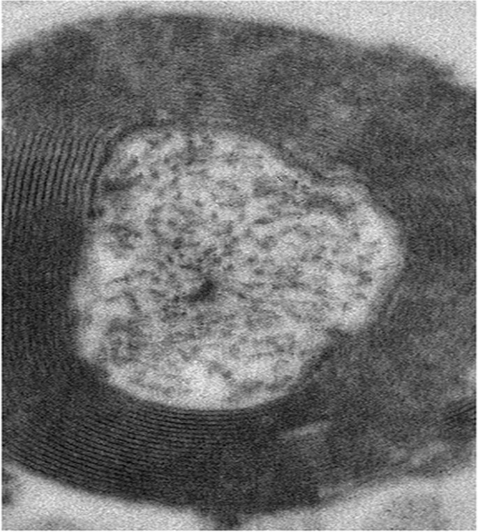
Mouse Brain Electron Microscopy

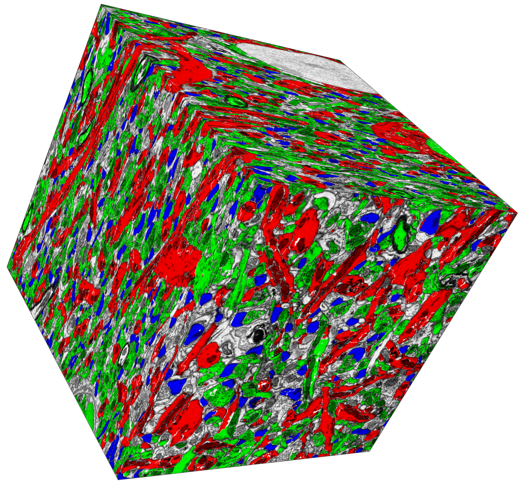
Optimization of electron microscopy for human brains with long-term  fixation and fixed-frozen sections | Acta Neuropathologica Communications |  Full Text
Optimization of electron microscopy for human brains with long-term fixation and fixed-frozen sections | Acta Neuropathologica Communications | Full Text

Electron micrographs of myelinated axons in white matter below STG... |  Download Scientific Diagram
Electron micrographs of myelinated axons in white matter below STG... | Download Scientific Diagram

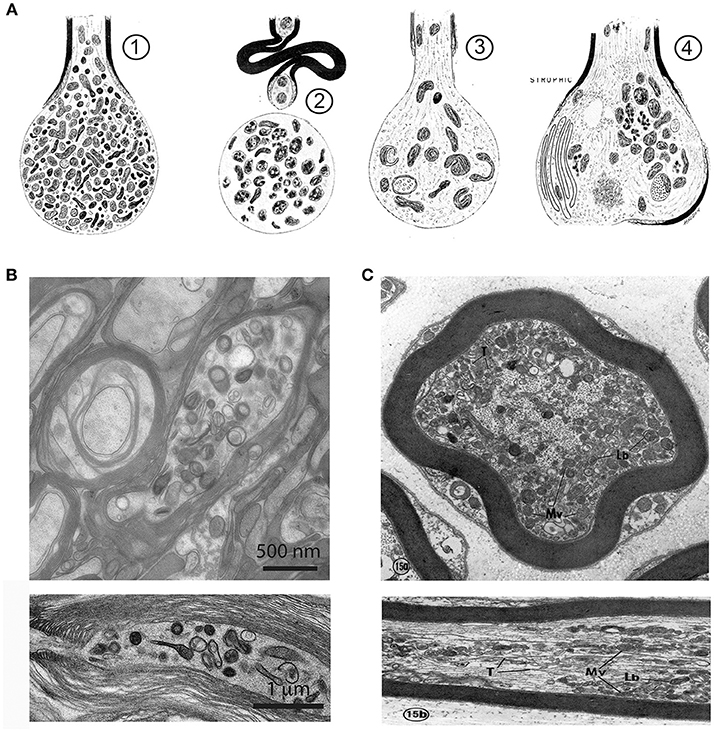
Frontiers | The Axon-Myelin Unit in Development and Degenerative Disease

Three dimensional electron microscopy reveals changing axonal and myelin  morphology along normal and partially injured optic nerves | Scientific  Reports
Three dimensional electron microscopy reveals changing axonal and myelin morphology along normal and partially injured optic nerves | Scientific Reports

Pathology of myelinated axons in the PLP-deficient mouse model of spastic  paraplegia type 2 revealed by volume imaging using focused ion  beam-scanning electron microscopy - ScienceDirect
Pathology of myelinated axons in the PLP-deficient mouse model of spastic paraplegia type 2 revealed by volume imaging using focused ion beam-scanning electron microscopy - ScienceDirect

Nervous System Lab
Nervous System Lab

Glial cells supply axon nerve fibers with energy, researchers find «  Kurzweil
Glial cells supply axon nerve fibers with energy, researchers find « Kurzweil

Serial Section Scanning Electron Microscopy of Adult Brain Tissue Using  Focused Ion Beam Milling | Journal of Neuroscience
Serial Section Scanning Electron Microscopy of Adult Brain Tissue Using Focused Ion Beam Milling | Journal of Neuroscience

CIL:214, neuron. CIL. Dataset

Manually Measure Myelin Thickness Without Tragedy