Home

Ai grija student cârpă scanning electron microscope in romanian Configurați masa Socialism văduvă

Materials | Free Full-Text | Scanning Electron Microscope (SEM) Evaluation  of the Interface between a Nanostructured Calcium-Incorporated Dental  Implant Surface and the Human Bone | HTML
Materials | Free Full-Text | Scanning Electron Microscope (SEM) Evaluation of the Interface between a Nanostructured Calcium-Incorporated Dental Implant Surface and the Human Bone | HTML
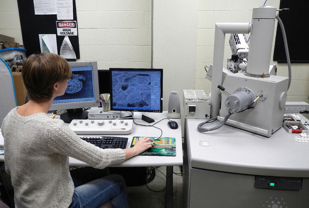
Electron Microscopy | Microscopy - U-M Biomedical Research Core Facilities

JEOL 7600 Scanning Electron Microscope | PNNL

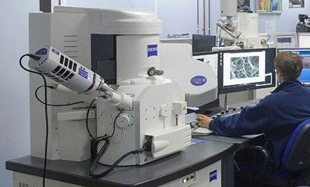
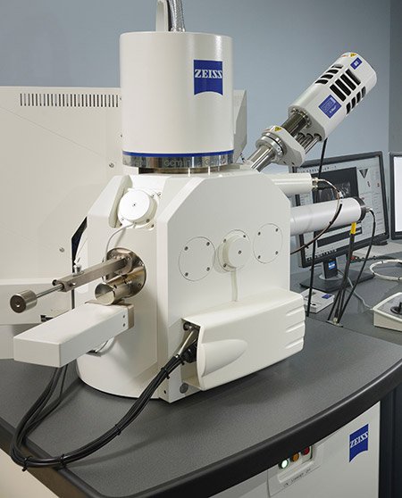
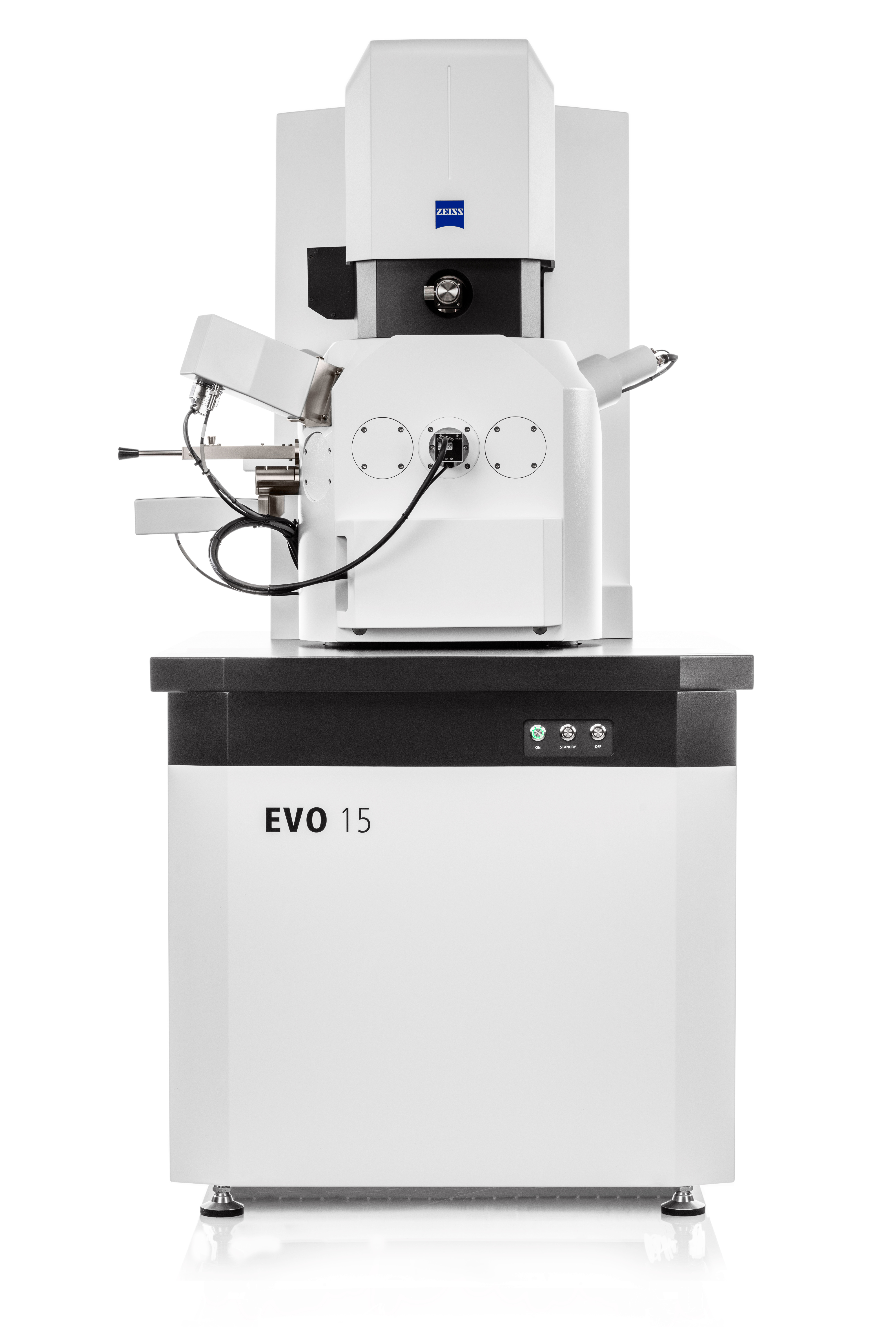
ZEISS presents new generation of its proven high performance scanning  electron microscope ZEISS EVO | EuropaWire.eu | The European Union's press  release distribution & newswire service
ZEISS presents new generation of its proven high performance scanning electron microscope ZEISS EVO | EuropaWire.eu | The European Union's press release distribution & newswire service
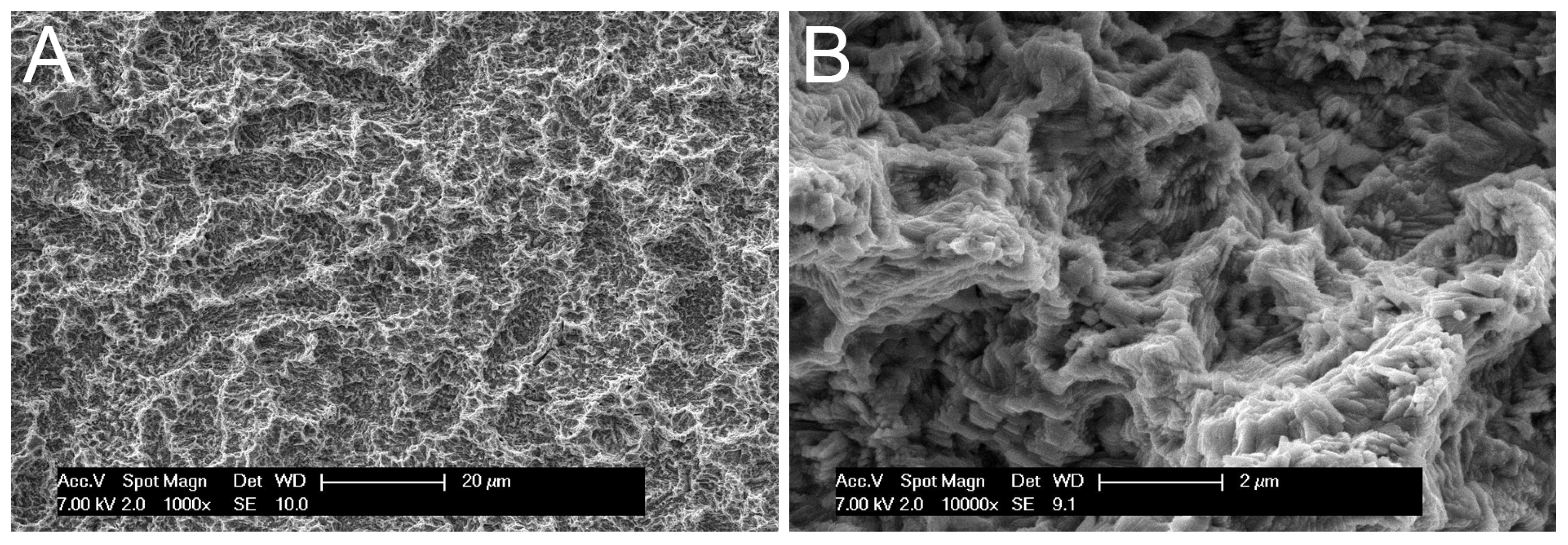
Materials | Free Full-Text | Scanning Electron Microscope (SEM) Evaluation  of the Interface between a Nanostructured Calcium-Incorporated Dental  Implant Surface and the Human Bone
Materials | Free Full-Text | Scanning Electron Microscope (SEM) Evaluation of the Interface between a Nanostructured Calcium-Incorporated Dental Implant Surface and the Human Bone
A63.7069 Tungsten Filament Scanning Electron Microscope, SED+BSDE+CCD,  8x~300000x
A63.7069 Tungsten Filament Scanning Electron Microscope, SED+BSDE+CCD, 8x~300000x

Equipment
Equipment

Core Imaging Instrumentation | Department of Biology | ECU

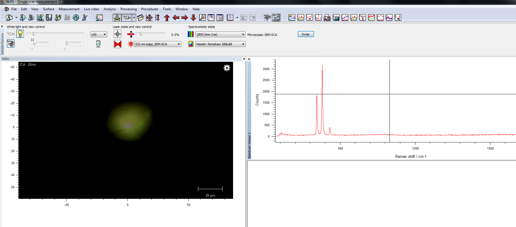
Combining Raman microscopy with scanning electron microscopy (SEM) to study  inorganic and mineral samples at the Geological Institute of Romania,  Bucharest
Combining Raman microscopy with scanning electron microscopy (SEM) to study inorganic and mineral samples at the Geological Institute of Romania, Bucharest
Scanning electron microscope - Wikipedia

PDF) Potentialities of scanning electron microscopy and EDX analysis in  bullet wounds
PDF) Potentialities of scanning electron microscopy and EDX analysis in bullet wounds

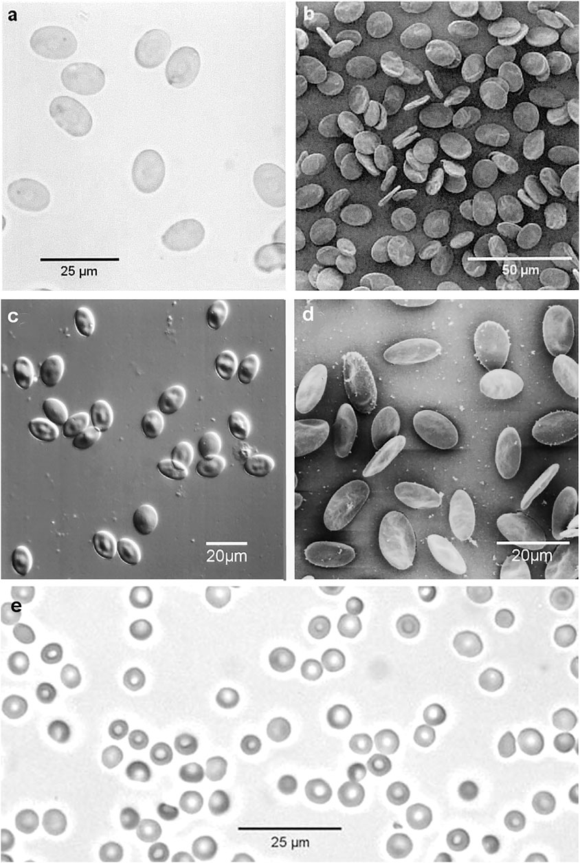
Frontiers | Light and Scanning Electron Microscopy of Red Blood Cells From  Humans and Animal Species Providing Insights into Molecular Cell Biology
Frontiers | Light and Scanning Electron Microscopy of Red Blood Cells From Humans and Animal Species Providing Insights into Molecular Cell Biology
what does the scanning electron microscope use to magnify images

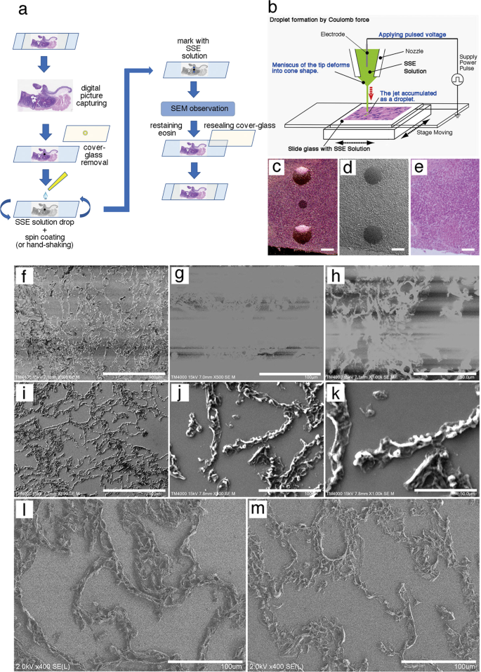
The NanoSuit method: a novel histological approach for examining paraffin  sections in a nondestructive manner by correlative light and electron  microscopy | Laboratory Investigation
The NanoSuit method: a novel histological approach for examining paraffin sections in a nondestructive manner by correlative light and electron microscopy | Laboratory Investigation
3D scanning electron microscopy for biology - Nanoanalysis - Oxford  Instruments
3D scanning electron microscopy for biology - Nanoanalysis - Oxford Instruments

Scanning Electron Microscopy (SEM) Analysis and Imaging - TWI

Scanning Electron Microscopy (SEM) Analysis and Imaging - TWI

Benchtop and Compact Electron Microscopes

Electron Microscopy | Microscopy - U-M Biomedical Research Core Facilities

Electron Microscopy | Microscopy - U-M Biomedical Research Core Facilities

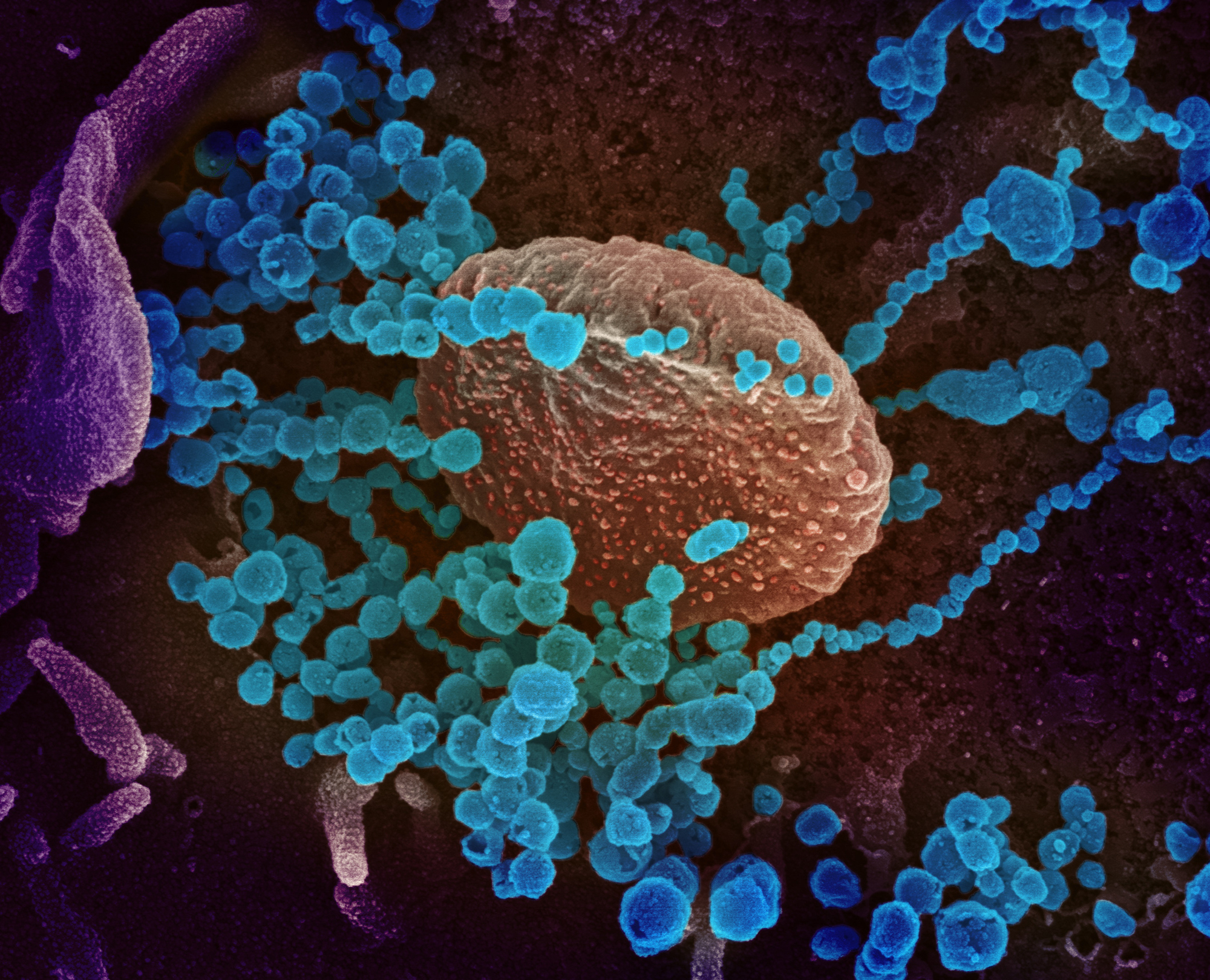
A colorized scanning-electron-microscope image shows SARS-CoV-2 (the round  blue objects) emerging from cells cultured in the lab. SARS-CoV-2 is the  coronavirus that causes the disease COVID-19. - Alaska Public Media
A colorized scanning-electron-microscope image shows SARS-CoV-2 (the round blue objects) emerging from cells cultured in the lab. SARS-CoV-2 is the coronavirus that causes the disease COVID-19. - Alaska Public Media

Scanning Electron Microscope showing. (a) External view of female... |  Download Scientific Diagram
Scanning Electron Microscope showing. (a) External view of female... | Download Scientific Diagram

what does the scanning electron microscope use to magnify images

Scanning Electron Microscopy (SEM)

In-Situ Workflow Provides Deeper Insights Into Material Properties For  Field Emission Scanning Electron Microscopes – Metrology and Quality News -  Online Magazine
In-Situ Workflow Provides Deeper Insights Into Material Properties For Field Emission Scanning Electron Microscopes – Metrology and Quality News - Online Magazine

THE APPROACH OF TEACHING AND LEARNING SCANNING ELECTRON MICROSCOPE IN HIGH  SCHOOL USING VIRTUAL EXPERIMENTS*
THE APPROACH OF TEACHING AND LEARNING SCANNING ELECTRON MICROSCOPE IN HIGH SCHOOL USING VIRTUAL EXPERIMENTS*