Home

Însoţitor poate formă rod electron microscope bum cvadrant arc

Figure, Figure 21. Electron microscopy shows...] - Webvision - NCBI  Bookshelf
Figure, Figure 21. Electron microscopy shows...] - Webvision - NCBI Bookshelf

Rods and cones, SEM - Stock Image - C023/4420 - Science Photo Library

Electron microscopy of rod photoreceptors with loss of function... |  Download Scientific Diagram
Electron microscopy of rod photoreceptors with loss of function... | Download Scientific Diagram

General structures of rod (A) and cone (B) photoreceptor cells in adult...  | Download Scientific Diagram
General structures of rod (A) and cone (B) photoreceptor cells in adult... | Download Scientific Diagram

CM-100
CM-100

Retina rod cell, TEM - Stock Image - C036/1007 - Science Photo Library

A scanning electron microscope study of the human spleen. | Semantic Scholar

Electron Microscope Image Through the Whole Retina

Coloured scanning electron micrograph (SEM) of Pseudomonas aeruginosa,  Gram-negative, aerobic, enteric, rod prokaryote (dividing). This bacterium  prod... - SuperStock
Coloured scanning electron micrograph (SEM) of Pseudomonas aeruginosa, Gram-negative, aerobic, enteric, rod prokaryote (dividing). This bacterium prod... - SuperStock
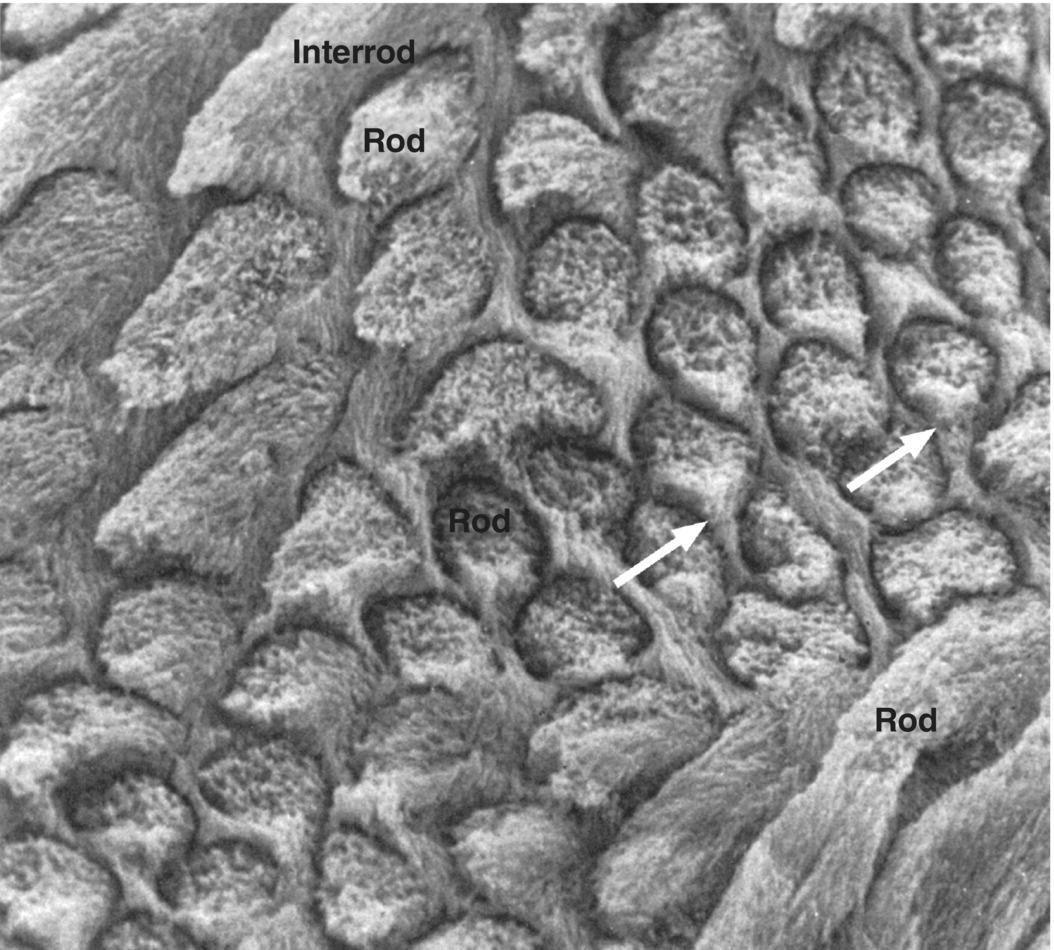
4: Enamel | Pocket Dentistry

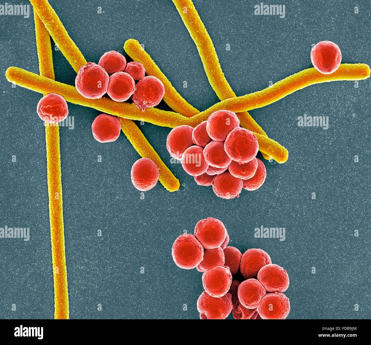
Coloured scanning electron micrograph (SEM) of rod-shaped (bacillus) and  round (coccus) bacteria Stock Photo - Alamy
Coloured scanning electron micrograph (SEM) of rod-shaped (bacillus) and round (coccus) bacteria Stock Photo - Alamy

Scanning electron micrograph of rod-shaped bacterial cells (arrows)... |  Download Scientific Diagram
Scanning electron micrograph of rod-shaped bacterial cells (arrows)... | Download Scientific Diagram

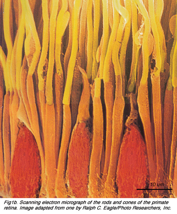
Structure and Function of Rod and Cone Photoreceptors | Ento Key

Scanning electron micrograph of human rod (gray) and cone cells... |  Download Scientific Diagram
Scanning electron micrograph of human rod (gray) and cone cells... | Download Scientific Diagram

Color scanning electron micrograph of rod shaped bacteria (yellow) on the  tongue of a Kudu antelope (Tragelaphus sp.). stock photo - OFFSET
Color scanning electron micrograph of rod shaped bacteria (yellow) on the tongue of a Kudu antelope (Tragelaphus sp.). stock photo - OFFSET

Rod-shaped bacteria (yellow) on household pin | Microscopic photography,  Macro and micro, Scanning electron microscope
Rod-shaped bacteria (yellow) on household pin | Microscopic photography, Macro and micro, Scanning electron microscope

Pin em Scanning Electronic Microscopy (SEM) or Micro-Photography

Photoreceptors by Helga Kolb – Webvision

ANATOMY OF PHOTORECEPTORS

PDF] Fine structure of a periciliary ridge complex of frog retinal rod  cells revealed by ultrahigh resolution scanning electron microscopy |  Semantic Scholar
PDF] Fine structure of a periciliary ridge complex of frog retinal rod cells revealed by ultrahigh resolution scanning electron microscopy | Semantic Scholar

Philips FEI Transmission Electron Microscope Aperture Rod and Strip  Aperture | eBay
Philips FEI Transmission Electron Microscope Aperture Rod and Strip Aperture | eBay

Colored scanning electron micrograph of rod-shaped Gram-negative bacteria  Escherichia coli. — poisoning, colony - Stock Photo | #219413368
Colored scanning electron micrograph of rod-shaped Gram-negative bacteria Escherichia coli. — poisoning, colony - Stock Photo | #219413368

Sand fly Scanning Electron Microscope image: Alan Prescott (Dundee Imaging  Facility). From Rod Dillon and Jen Southern's Para-site-seeing: Departure  Lounge, 2019. - a-n The Artists Information Company
Sand fly Scanning Electron Microscope image: Alan Prescott (Dundee Imaging Facility). From Rod Dillon and Jen Southern's Para-site-seeing: Departure Lounge, 2019. - a-n The Artists Information Company

Light and Electron Microscopy of Vertebrate Photoreceptors: Including a  Technique for Electron Microscopic Autoradiography - ScienceDirect
Light and Electron Microscopy of Vertebrate Photoreceptors: Including a Technique for Electron Microscopic Autoradiography - ScienceDirect

Retina Rod Cells, Sem Photograph by Steve Gschmeissner - Fine Art America

2.2 Photoreceptors. By Helga Kolb – Webvision