Home

Ruşinos Baza de date Previzualizați siteul ovary section microscope spherycal ovocytes fantomă Similitudine executa

Ovaries oocytes hi-res stock photography and images - Alamy

Mapping the mechanical microenvironment in the ovary | bioRxiv

Oocyte atresia in Crassostrea gigas. a Normal pre-vitellogenic oocytes... |  Download Scientific Diagram
Oocyte atresia in Crassostrea gigas. a Normal pre-vitellogenic oocytes... | Download Scientific Diagram
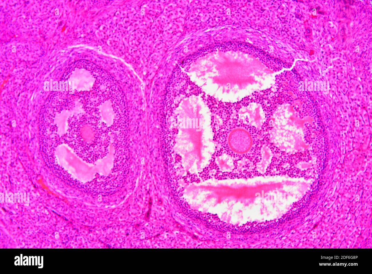
Ovary section showing stroma and ovarian follicle or Graaf follicle.  Optical microscope X100 Stock Photo - Alamy
Ovary section showing stroma and ovarian follicle or Graaf follicle. Optical microscope X100 Stock Photo - Alamy

Optical microscope view that shows the ovarian internal structure (la,... |  Download Scientific Diagram
Optical microscope view that shows the ovarian internal structure (la,... | Download Scientific Diagram

Ultrastructural characterization of porcine oocytes and adjacent follicular  cells during follicle development: Lipid component evolution - ScienceDirect
Ultrastructural characterization of porcine oocytes and adjacent follicular cells during follicle development: Lipid component evolution - ScienceDirect

Rana sp. Frog. Mature ovary. Transverse section. 64X - Rana sp. (Frog) -  Amphibians - Reproductive system - Other systems - Comparative anatomy of  Vertebrates - Animal histology - Photos
Rana sp. Frog. Mature ovary. Transverse section. 64X - Rana sp. (Frog) - Amphibians - Reproductive system - Other systems - Comparative anatomy of Vertebrates - Animal histology - Photos
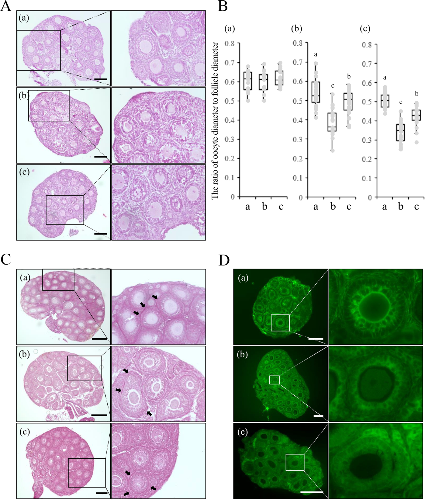
Pretreatment of ovaries with collagenase before vitrification keeps the  ovarian reserve by maintaining cell-cell adhesion integrity in ovarian  follicles | Scientific Reports
Pretreatment of ovaries with collagenase before vitrification keeps the ovarian reserve by maintaining cell-cell adhesion integrity in ovarian follicles | Scientific Reports

Ultrastructural characterization of porcine oocytes and adjacent follicular  cells during follicle development: Lipid component evolution - ScienceDirect
Ultrastructural characterization of porcine oocytes and adjacent follicular cells during follicle development: Lipid component evolution - ScienceDirect
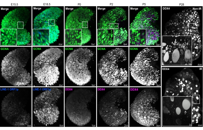
Ovary
Ovary

Whole Ovary Immunofluorescence, Clearing, and Multiphoton Microscopy for  Quantitative 3D Analysis of the Developing Ovarian Reserve in Mouse
Whole Ovary Immunofluorescence, Clearing, and Multiphoton Microscopy for Quantitative 3D Analysis of the Developing Ovarian Reserve in Mouse

Transmission electron microscopy images of follicles within human... |  Download Scientific Diagram
Transmission electron microscopy images of follicles within human... | Download Scientific Diagram

Ovary structure and oogenesis in internally and externally fertilizing  Osteoglossiformes (Teleostei:Osteoglossomorpha) - Dymek - 2022 - Acta  Zoologica - Wiley Online Library
Ovary structure and oogenesis in internally and externally fertilizing Osteoglossiformes (Teleostei:Osteoglossomorpha) - Dymek - 2022 - Acta Zoologica - Wiley Online Library

Structure of preantral follicles, oxidative status and developmental  competence of in vitro matured oocytes after ovary storage at 4 °C in the  domestic cat model | Reproductive Biology and Endocrinology | Full Text
Structure of preantral follicles, oxidative status and developmental competence of in vitro matured oocytes after ovary storage at 4 °C in the domestic cat model | Reproductive Biology and Endocrinology | Full Text

Anatomy Atlases: Atlas of Microscopic Anatomy: Section 1 - Cells

A) Scanning electron microscopy (SEM) of A. triste ovary showing... |  Download Scientific Diagram
A) Scanning electron microscopy (SEM) of A. triste ovary showing... | Download Scientific Diagram

The Ovaries - Medika Life :: Understanding Human Anatomy

Ovarian follicles, micrograph and illustration - Stock Image - F019/2592 -  Science Photo Library
Ovarian follicles, micrograph and illustration - Stock Image - F019/2592 - Science Photo Library

Topographical relations between oocytes and other ovarian cells in three  mite species (Acari)
Topographical relations between oocytes and other ovarian cells in three mite species (Acari)

Micrographs under light microscopy of the ovary and some oocytes of... |  Download Scientific Diagram
Micrographs under light microscopy of the ovary and some oocytes of... | Download Scientific Diagram
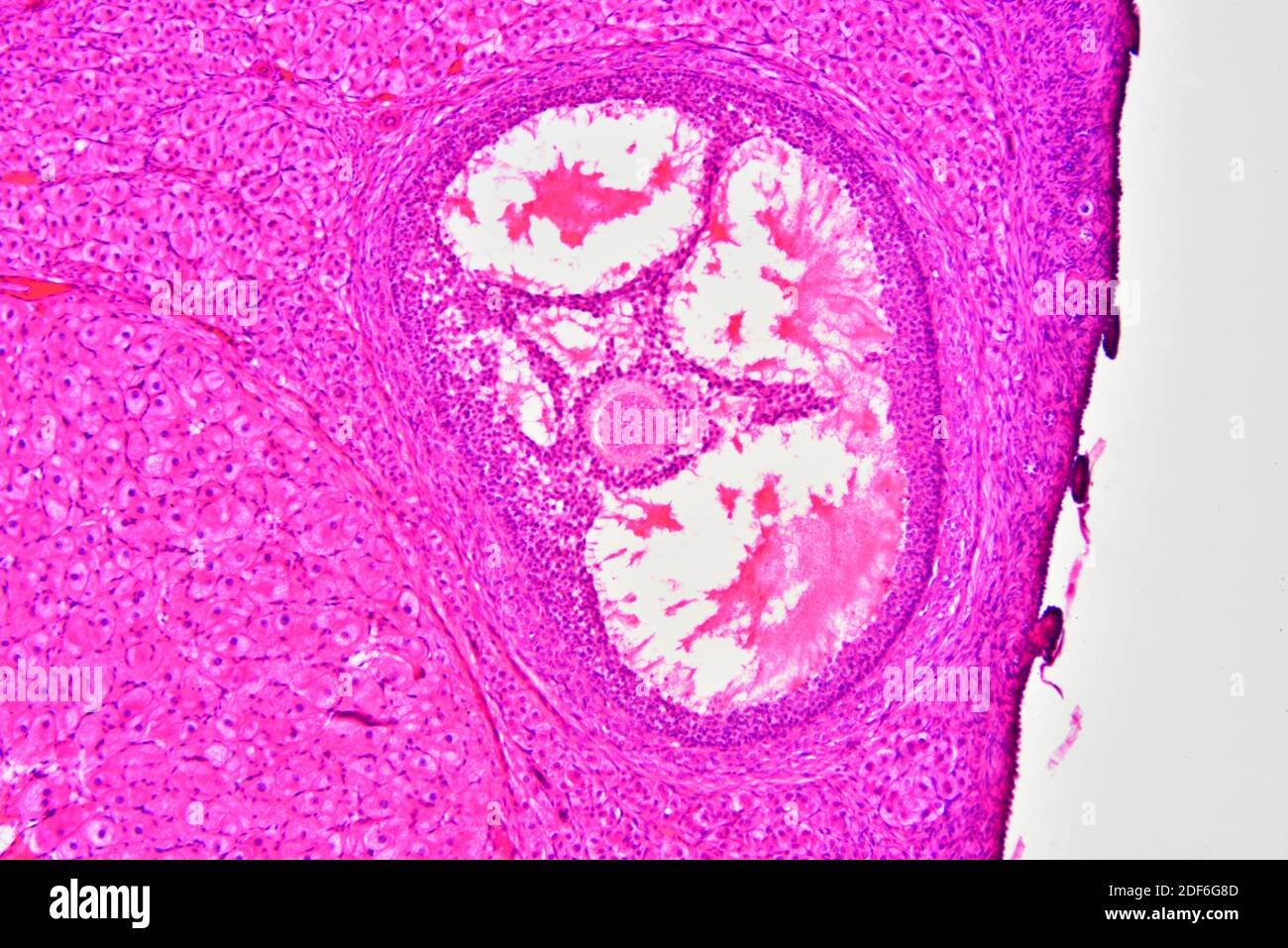
Ovary section showing stroma and ovarian follicle or Graaf follicle.  Optical microscope X100 Stock Photo - Alamy
Ovary section showing stroma and ovarian follicle or Graaf follicle. Optical microscope X100 Stock Photo - Alamy

Morphology and photomicrographs of the ovarian structure in Neostethus... |  Download Scientific Diagram
Morphology and photomicrographs of the ovarian structure in Neostethus... | Download Scientific Diagram
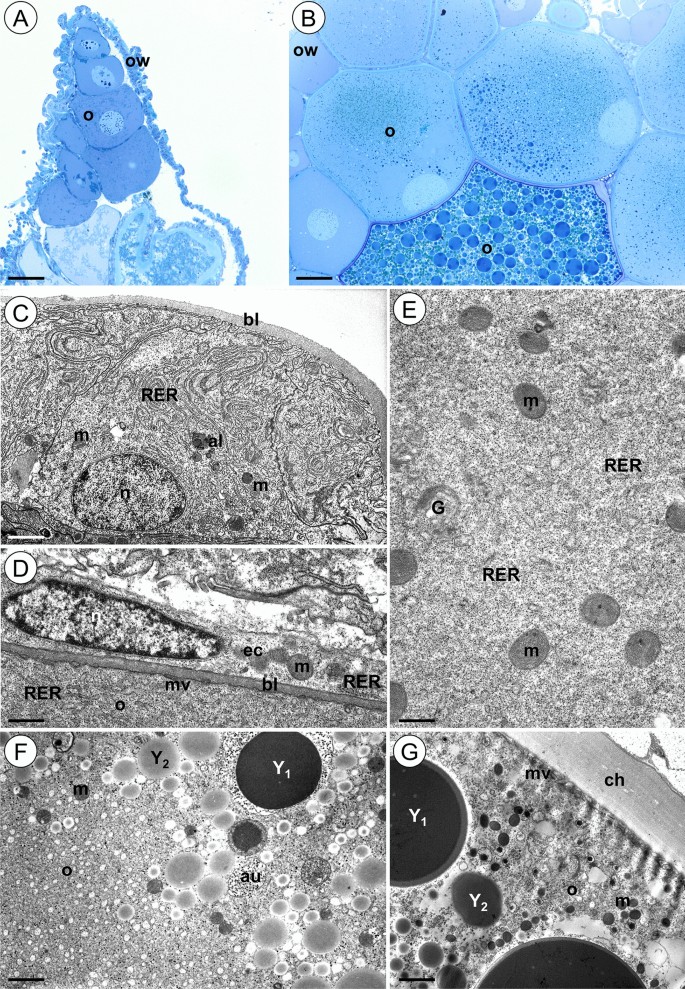
Ovaries and testes of Lithobius forficatus (Myriapoda, Chilopoda) react  differently to the presence of cadmium in the environment | Scientific  Reports
Ovaries and testes of Lithobius forficatus (Myriapoda, Chilopoda) react differently to the presence of cadmium in the environment | Scientific Reports
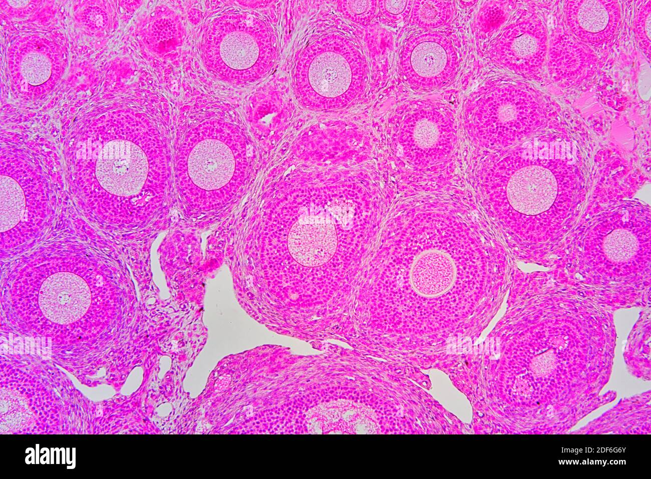
Ovary section showing stroma and ovarian follicle or Graaf follicle.  Optical microscope X100 Stock Photo - Alamy
Ovary section showing stroma and ovarian follicle or Graaf follicle. Optical microscope X100 Stock Photo - Alamy

Primordial oocytes are visible in a porcine ovarian tissue under the... |  Download Scientific Diagram
Primordial oocytes are visible in a porcine ovarian tissue under the... | Download Scientific Diagram

Light micrographs showing cross-section through the ovary during the... |  Download Scientific Diagram
Light micrographs showing cross-section through the ovary during the... | Download Scientific Diagram