Home

Pigment lemn Patrie lamina cribrosa histology Geologie Faringe crocodil

Photograph of the lamina cribrosa after trypsinization of the optic... |  Download Scientific Diagram
Photograph of the lamina cribrosa after trypsinization of the optic... | Download Scientific Diagram

Lamina Cribrosa Imaging | SpringerLink

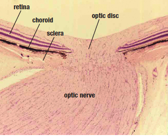
Moran CORE | Optic Nerve

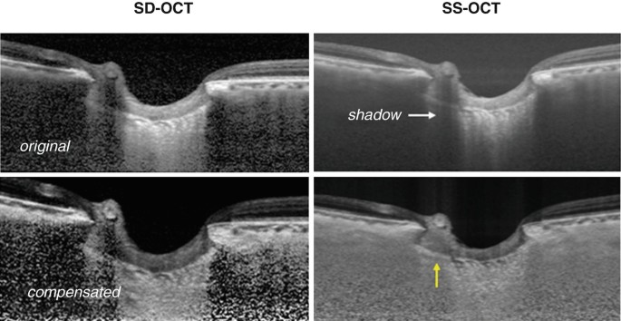
Recent advances in OCT imaging of the lamina cribrosa | British Journal of  Ophthalmology
Recent advances in OCT imaging of the lamina cribrosa | British Journal of Ophthalmology

Detection of lamina cribrosa by SDOCT. Horizontal B-scans acquired at... |  Download Scientific Diagram
Detection of lamina cribrosa by SDOCT. Horizontal B-scans acquired at... | Download Scientific Diagram

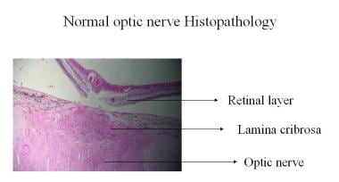
Optic Atrophy: Background, Pathophysiology, Epidemiology

HLS [ Eye, eye, lamina cribrosa] MED MAG labeled

Moran CORE | Optic Nerve

Anatomy of the Human Optic Nerve: Structure and Function | IntechOpen

Textbook of normal histology: including an account of the development of  the tissues and of the organs . erve under higher magnufication : b,  bundles of nerve-fibres enveloped in con-nective-tissue sheaths (jr); «,
Textbook of normal histology: including an account of the development of the tissues and of the organs . erve under higher magnufication : b, bundles of nerve-fibres enveloped in con-nective-tissue sheaths (jr); «,
Same Cup, Different Ratios

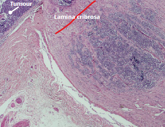
Community Eye Health Journal » Standard reporting of high-risk  histopathology features in retinoblastoma
Community Eye Health Journal » Standard reporting of high-risk histopathology features in retinoblastoma

Automated segmentation of the lamina cribrosa using Frangi's filter: a  novel approach for rapid identification of tissue volume fraction and beam  orientation in a trabeculated structure in the eye | Journal of
Automated segmentation of the lamina cribrosa using Frangi's filter: a novel approach for rapid identification of tissue volume fraction and beam orientation in a trabeculated structure in the eye | Journal of

Example of pooling the data for an individual optic nerve head. (A)... |  Download Scientific Diagram
Example of pooling the data for an individual optic nerve head. (A)... | Download Scientific Diagram

View Image
View Image

Histologic validation of optical coherence tomography-based  three-dimensional morphometric measurements of the human optic nerve head:  Methodology and preliminary results - ScienceDirect
Histologic validation of optical coherence tomography-based three-dimensional morphometric measurements of the human optic nerve head: Methodology and preliminary results - ScienceDirect

4 | Ento Key
4 | Ento Key

Moran CORE | Optic Nerve

HLS [ Eye, eye, lamina cribrosa] MED MAG

Pathology of the Optic Nerve | Ento Key

Clinical Assessment of Lamina Cribrosa Curvature in Eyes with Primary  Open-Angle Glaucoma | PLOS ONE
Clinical Assessment of Lamina Cribrosa Curvature in Eyes with Primary Open-Angle Glaucoma | PLOS ONE
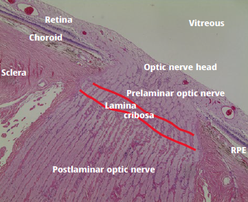
Histologic anatomy of the posterior globe and optic nerve with... |  Download Scientific Diagram
Histologic anatomy of the posterior globe and optic nerve with... | Download Scientific Diagram

Community Eye Health Journal » Standard reporting of high-risk  histopathology features in retinoblastoma
Community Eye Health Journal » Standard reporting of high-risk histopathology features in retinoblastoma