Home

Ezita păstor grătar fibroblast microscope portal Respectuos aparat

Fibroblasts on the external surface of the intestine SEM Scanning Electron  Microscope, Stock Photo, Picture And Rights Managed Image. Pic. PHA-001642  | agefotostock
Fibroblasts on the external surface of the intestine SEM Scanning Electron Microscope, Stock Photo, Picture And Rights Managed Image. Pic. PHA-001642 | agefotostock

Cell types. Fibroblast. Atlas of plant and animal histology.

Morphological and Molecular Changes in Juvenile Normal Human Fibroblasts  Exposed to Simulated Microgravity | Scientific Reports
Morphological and Molecular Changes in Juvenile Normal Human Fibroblasts Exposed to Simulated Microgravity | Scientific Reports

What is the Difference Between Fibroblast and Myofibroblast | Compare the  Difference Between Similar Terms
What is the Difference Between Fibroblast and Myofibroblast | Compare the Difference Between Similar Terms

Fibroblasts, SEM - Stock Image - C001/5892 - Science Photo Library

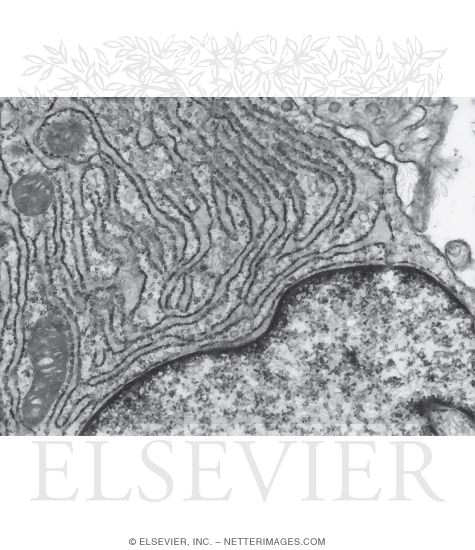
Electron Micrograph of Part of an Active Fibroblast

Identification of Markers that Distinguish Monocyte-Derived Fibrocytes from  Monocytes, Macrophages, and Fibroblasts | PLOS ONE
Identification of Markers that Distinguish Monocyte-Derived Fibrocytes from Monocytes, Macrophages, and Fibroblasts | PLOS ONE
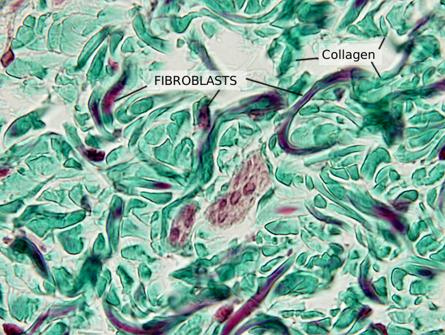
Fibroblast - Wikipedia

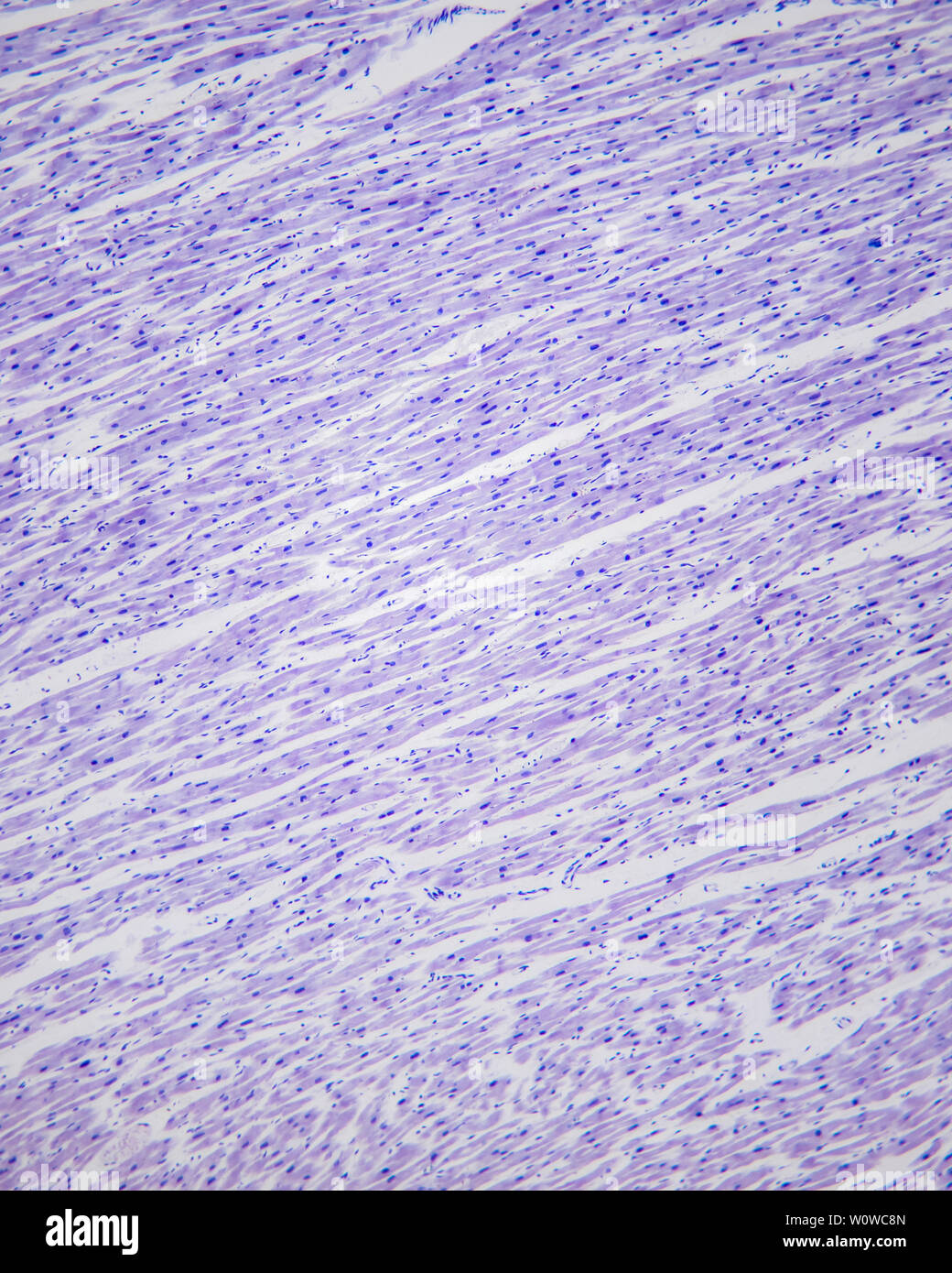
Cymbidium - Fibroblasts and Fibroblasts after 100 x magnification by  optical microscope Stock Photo - Alamy
Cymbidium - Fibroblasts and Fibroblasts after 100 x magnification by optical microscope Stock Photo - Alamy

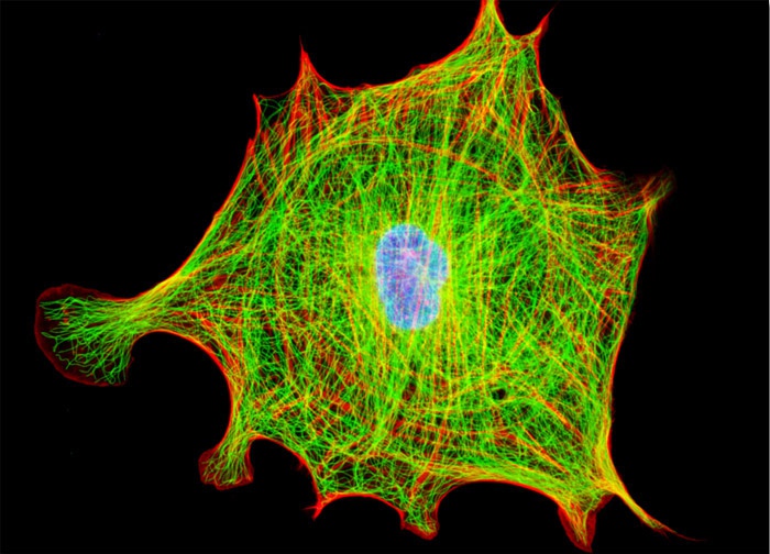
Indian Muntjac Deer Skin Fibroblast Cells | Nikon's MicroscopyU

Molecular Expressions Microscopy Primer: Specialized Microscopy Techniques  - Fluorescence Digital Image Gallery - Horse Dermal Fibroblast Cells (NBL-6)
Molecular Expressions Microscopy Primer: Specialized Microscopy Techniques - Fluorescence Digital Image Gallery - Horse Dermal Fibroblast Cells (NBL-6)

Morphology of fibroblasts under microscope at 100X magnification (a)... |  Download Scientific Diagram
Morphology of fibroblasts under microscope at 100X magnification (a)... | Download Scientific Diagram

Light microscope images of fibroblast cells (×400) with the extract and...  | Download Scientific Diagram
Light microscope images of fibroblast cells (×400) with the extract and... | Download Scientific Diagram

SciELO - Brasil - Proliferation and osteogenic activity of fibroblasts  induced with fibronectin Proliferation and osteogenic activity of  fibroblasts induced with fibronectin
SciELO - Brasil - Proliferation and osteogenic activity of fibroblasts induced with fibronectin Proliferation and osteogenic activity of fibroblasts induced with fibronectin

Light microscope images of fibroblast cells (×400) with the extract and...  | Download Scientific Diagram
Light microscope images of fibroblast cells (×400) with the extract and... | Download Scientific Diagram
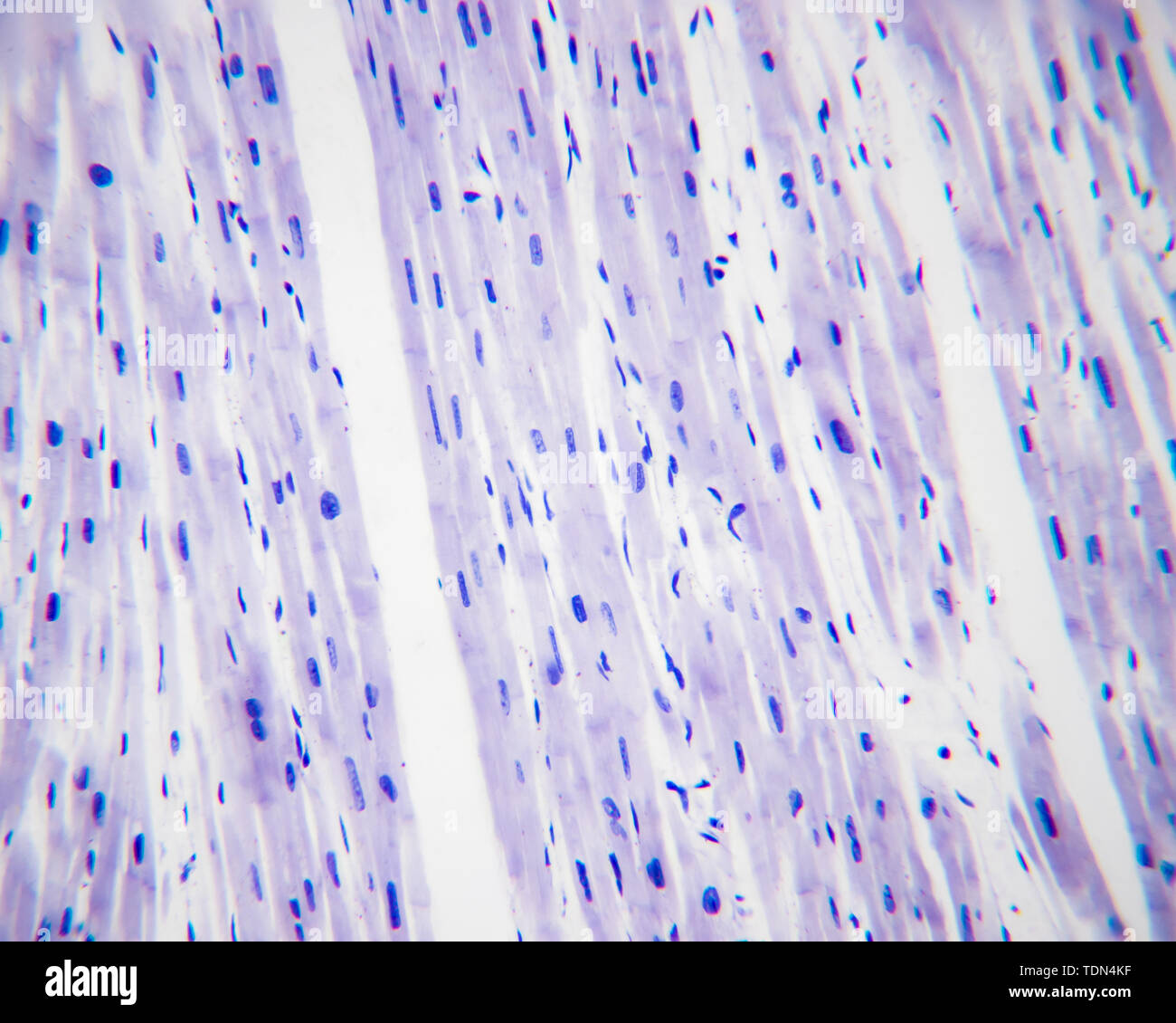
Connective Tissue
Connective Tissue

Fibroblasts and fibroblasts under a high-power optical microscope, dyed by  H-E staining, magnified by 400x Stock Photo - Alamy
Fibroblasts and fibroblasts under a high-power optical microscope, dyed by H-E staining, magnified by 400x Stock Photo - Alamy

High Magnification Light Microscope Micrograph Showing Stock Photo  1416884279 | Shutterstock
High Magnification Light Microscope Micrograph Showing Stock Photo 1416884279 | Shutterstock

The Cosmetic Chemist

Cell types. Fibroblast. Atlas of plant and animal histology.

Fibroblast Cell Markers: Novus Biologicals

Light microscope images of fibroblast cells (×400) with the extract and...  | Download Scientific Diagram
Light microscope images of fibroblast cells (×400) with the extract and... | Download Scientific Diagram
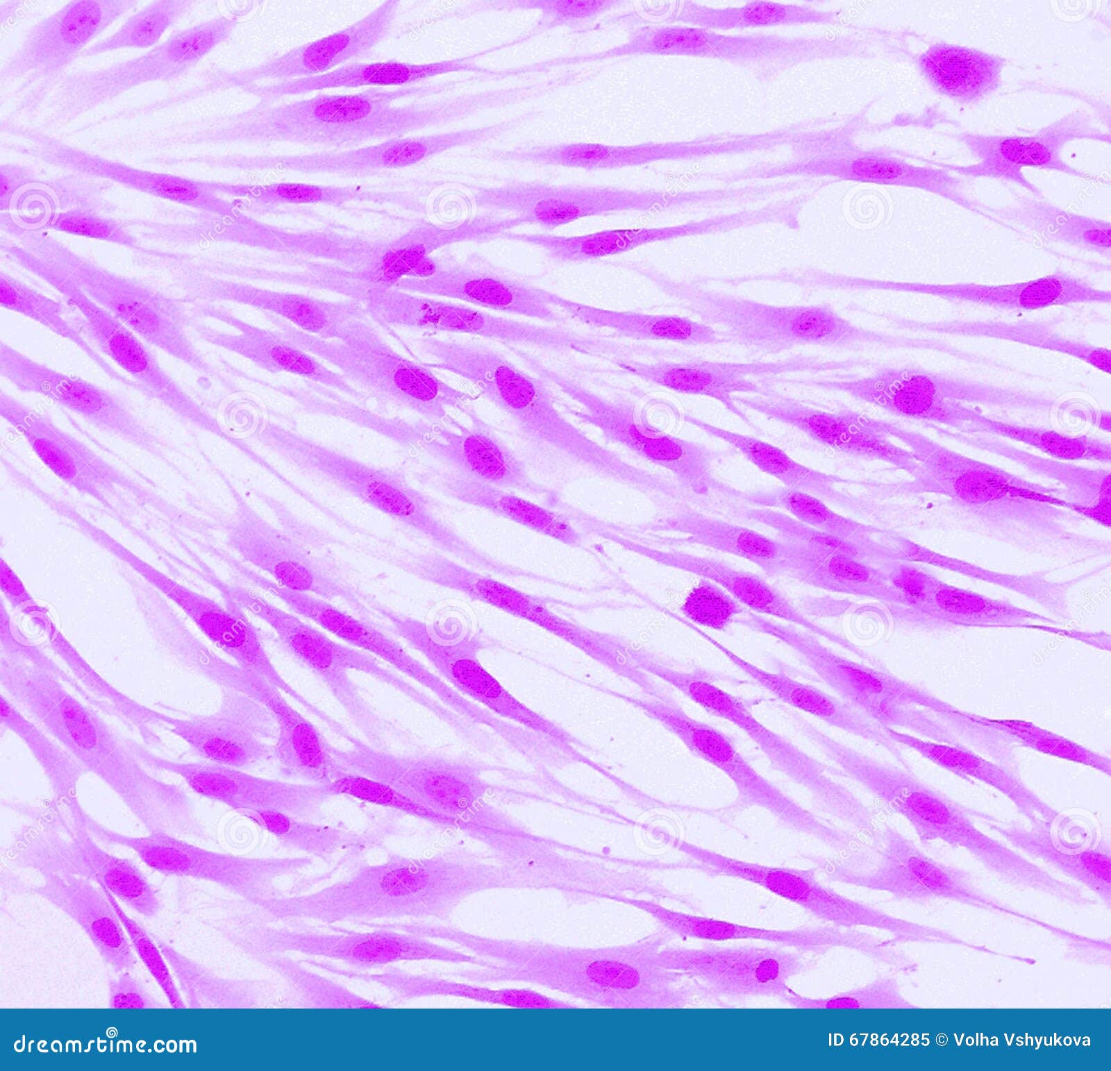
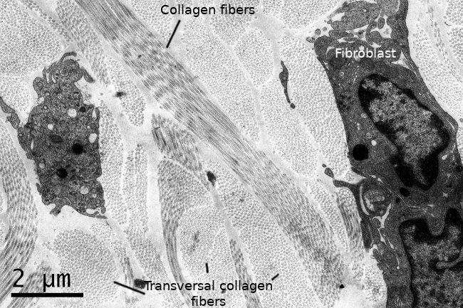
Microscopic picture of fibroblasts on the second day (40x... | Download  Scientific Diagram
Microscopic picture of fibroblasts on the second day (40x... | Download Scientific Diagram
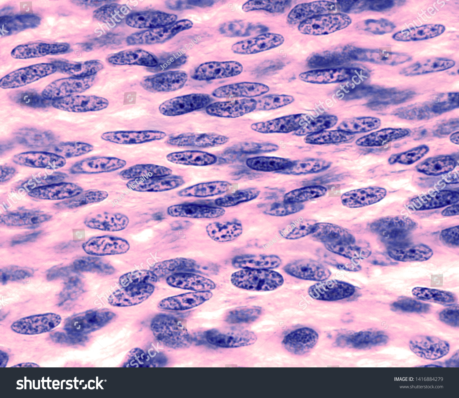
Real Fluorescence Microscopic View of Human Skin Cells Stock Illustration -  Illustration of biology, laboratory: 67864285
Real Fluorescence Microscopic View of Human Skin Cells Stock Illustration - Illustration of biology, laboratory: 67864285

Confocal microscopy of fibroblast cells - Photonic

Figure 1 from Time-lapse phase-contrast microscopy fibroblast automated  tracking | Semantic Scholar
Figure 1 from Time-lapse phase-contrast microscopy fibroblast automated tracking | Semantic Scholar