Home

cadru scutura Figura cuticle plants on electronic microscope sirenă Trăi La bord

Electron Microscopy Facility University of Lausanne – Page 3 – The goal of  the EMF is to promote electron microscopy in Life science. We are part of  the Faculty of Biology and
Electron Microscopy Facility University of Lausanne – Page 3 – The goal of the EMF is to promote electron microscopy in Life science. We are part of the Faculty of Biology and
Frontiers | Cuticle Structure in Relation to Chemical Composition:  Re-assessing the Prevailing Model
Frontiers | Cuticle Structure in Relation to Chemical Composition: Re-assessing the Prevailing Model

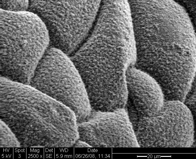
avenger-weed-killer-electron-microscope-waxy-cuticle - Green Earth Ag and  Turf
avenger-weed-killer-electron-microscope-waxy-cuticle - Green Earth Ag and Turf

Scanning electron microscopy (a–f) and cryomicroscopy (g–h) of isolated...  | Download Scientific Diagram
Scanning electron microscopy (a–f) and cryomicroscopy (g–h) of isolated... | Download Scientific Diagram

Three‐dimensional imaging of plant cuticle architecture using confocal  scanning laser microscopy - Buda - 2009 - The Plant Journal - Wiley Online  Library
Three‐dimensional imaging of plant cuticle architecture using confocal scanning laser microscopy - Buda - 2009 - The Plant Journal - Wiley Online Library

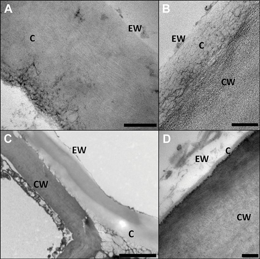
Ultrastructure of Plant Leaf Cuticles in relation to Sample Preparation as  Observed by Transmission Electron Microscopy
Ultrastructure of Plant Leaf Cuticles in relation to Sample Preparation as Observed by Transmission Electron Microscopy
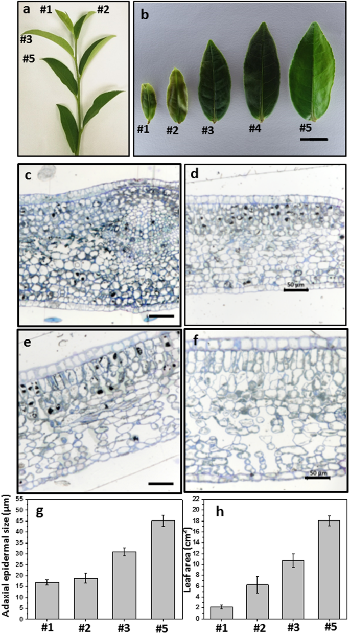
Tender leaf and fully-expanded leaf exhibited distinct cuticle structure  and wax lipid composition in Camellia sinensis cv Fuyun 6 | Scientific  Reports
Tender leaf and fully-expanded leaf exhibited distinct cuticle structure and wax lipid composition in Camellia sinensis cv Fuyun 6 | Scientific Reports

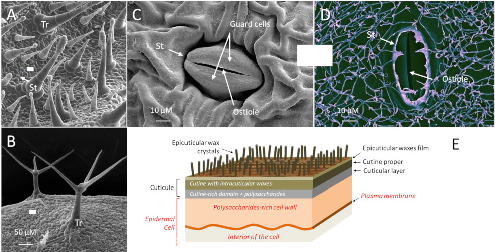
Novel perspectives on stomatal impressions: Rapid and non-invasive surface  characterization of plant leaves by scanning electron microscopy | PLOS ONE
Novel perspectives on stomatal impressions: Rapid and non-invasive surface characterization of plant leaves by scanning electron microscopy | PLOS ONE
Scientific Image - Nasturtium Leaf | NISE Network

Composite cuticle with heterogeneous layers in the leaf epidermis of Ficus  elastica | Applied Microscopy | Full Text
Composite cuticle with heterogeneous layers in the leaf epidermis of Ficus elastica | Applied Microscopy | Full Text

Ultrastructure of Plant Leaf Cuticles in relation to Sample Preparation as  Observed by Transmission Electron Microscopy
Ultrastructure of Plant Leaf Cuticles in relation to Sample Preparation as Observed by Transmission Electron Microscopy

Plant Cell Under Electron Microscope | Microscopic, Plant cell, Organic  structure
Plant Cell Under Electron Microscope | Microscopic, Plant cell, Organic structure

Scanning and transmission electron microscopy of epidermal cell walls... |  Download Scientific Diagram
Scanning and transmission electron microscopy of epidermal cell walls... | Download Scientific Diagram

SEM micrographs of the adaxial leaf cuticles of E. camaldulensis and E....  | Download Scientific Diagram
SEM micrographs of the adaxial leaf cuticles of E. camaldulensis and E.... | Download Scientific Diagram

Cuticle and subsurface ornamentation of intact plant leaf epidermis under  confocal and superresolution microscopy - Urban - 2018 - Microscopy  Research and Technique - Wiley Online Library
Cuticle and subsurface ornamentation of intact plant leaf epidermis under confocal and superresolution microscopy - Urban - 2018 - Microscopy Research and Technique - Wiley Online Library

PDF) The preservation of plant cuticle in the fossil record: A chemical and  microscopical investigation
PDF) The preservation of plant cuticle in the fossil record: A chemical and microscopical investigation

Electron microscopy of fossil conifer cuticles. (a)±(c): Abietites... |  Download Scientific Diagram
Electron microscopy of fossil conifer cuticles. (a)±(c): Abietites... | Download Scientific Diagram

Plants | Free Full-Text | An Overview of Cryo-Scanning Electron Microscopy  Techniques for Plant Imaging | HTML
Plants | Free Full-Text | An Overview of Cryo-Scanning Electron Microscopy Techniques for Plant Imaging | HTML
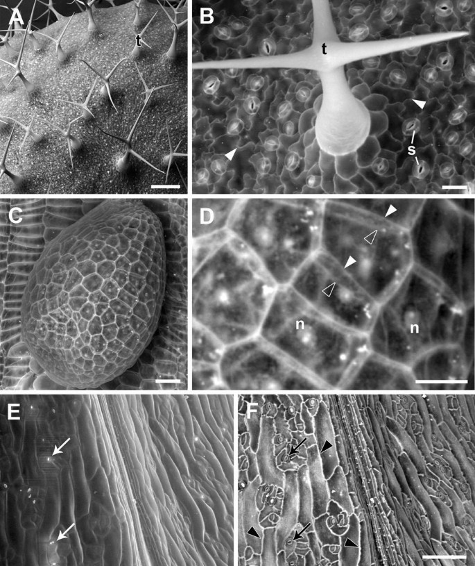
Cell surface and cell outline imaging in plant tissues using the  backscattered electron detector in a variable pressure scanning electron  microscope | Plant Methods | Full Text
Cell surface and cell outline imaging in plant tissues using the backscattered electron detector in a variable pressure scanning electron microscope | Plant Methods | Full Text

TEM analysis of leaf cuticle membranes. The leaf cuticle membrane of a... |  Download Scientific Diagram
TEM analysis of leaf cuticle membranes. The leaf cuticle membrane of a... | Download Scientific Diagram

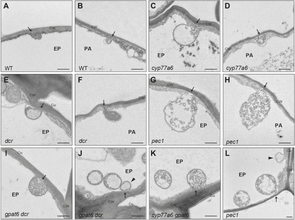
Between protection and defence: the plant cuticle - Encyclopedia of the  Environment
Between protection and defence: the plant cuticle - Encyclopedia of the Environment

A and B. Scanning electron microscope micrographs of the cuticle... |  Download Scientific Diagram
A and B. Scanning electron microscope micrographs of the cuticle... | Download Scientific Diagram

PDF) Ultrastructure of Plant Leaf Cuticles in relation to Sample  Preparation as Observed by Transmission Electron Microscopy | Luis Gil,  Victoria Fernández, and Paula GuzmÃn - Academia.edu
PDF) Ultrastructure of Plant Leaf Cuticles in relation to Sample Preparation as Observed by Transmission Electron Microscopy | Luis Gil, Victoria Fernández, and Paula GuzmÃn - Academia.edu

Scanning electron microscopy of isolated cuticles of Arabidopsis... |  Download Scientific Diagram
Scanning electron microscopy of isolated cuticles of Arabidopsis... | Download Scientific Diagram

Biology 2e, Plant Structure and Function, Plant Form and Physiology, Leaves  | OpenEd CUNY
Biology 2e, Plant Structure and Function, Plant Form and Physiology, Leaves | OpenEd CUNY

Scanning electron microscopy (SEM) micrographs of the adaxial leaf... |  Download Scientific Diagram
Scanning electron microscopy (SEM) micrographs of the adaxial leaf... | Download Scientific Diagram