Home

director Biblioteca trunchiului adăpost colors of fluorescence markers microscopy Caligrafie cocaină Comportament

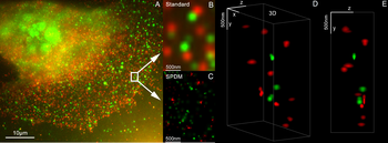
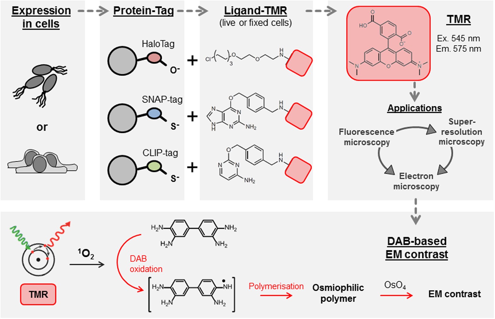
Self-labelling enzymes as universal tags for fluorescence microscopy,  super-resolution microscopy and electron microscopy | Scientific Reports
Self-labelling enzymes as universal tags for fluorescence microscopy, super-resolution microscopy and electron microscopy | Scientific Reports
2-color Fluorescence Microscopy System – Doric Lenses Inc.

Molecules | Free Full-Text | Advanced Fluorescence Microscopy  Techniques—FRAP, FLIP, FLAP, FRET and FLIM | HTML
Molecules | Free Full-Text | Advanced Fluorescence Microscopy Techniques—FRAP, FLIP, FLAP, FRET and FLIM | HTML

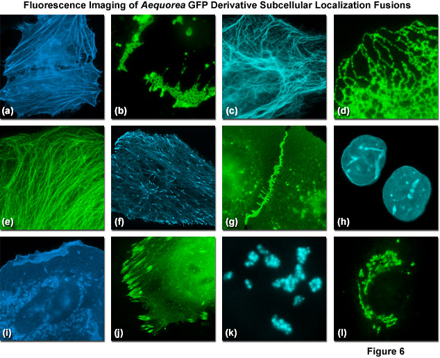
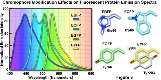
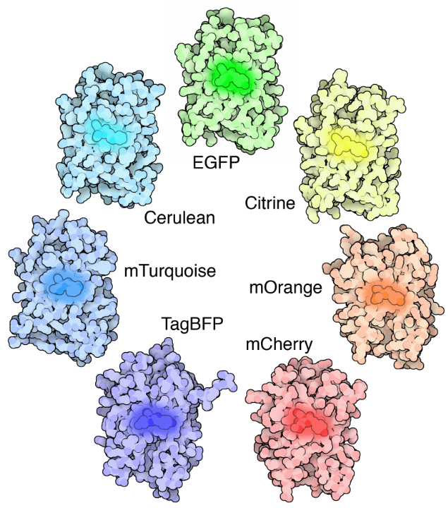
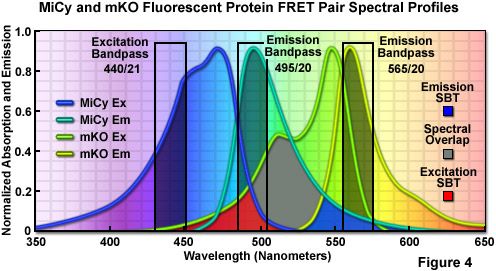
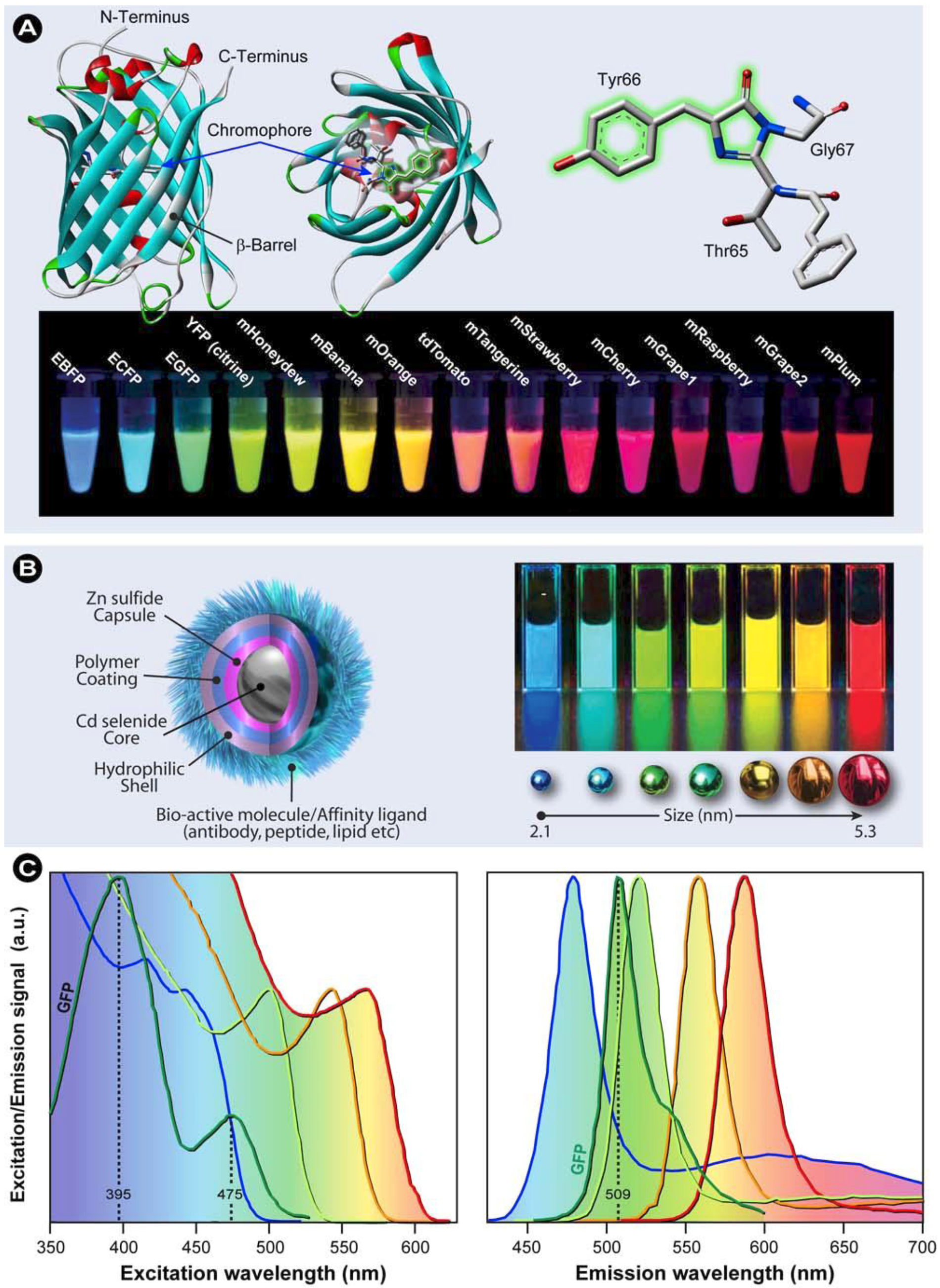
ZEISS Microscopy Online Campus | Jellyfish Fluorescent Proteins

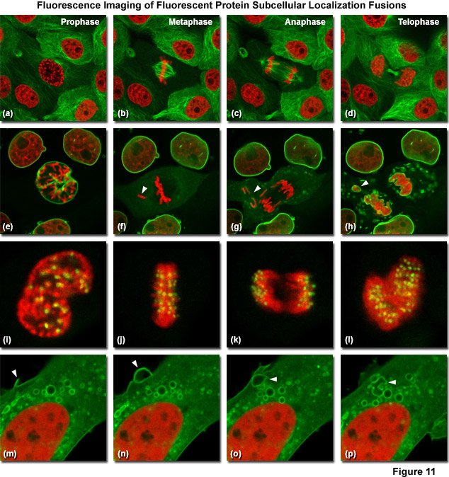
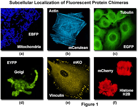
ZEISS Microscopy Online Campus | Introduction to Fluorescent Proteins

Fluorescence microscope - Wikipedia

Importance of Colocalization Studies

Fluorescence microscope - Wikipedia

A photostable fluorescent marker for the superresolution live imaging of  the dynamic structure of the mitochondrial cristae | PNAS
A photostable fluorescent marker for the superresolution live imaging of the dynamic structure of the mitochondrial cristae | PNAS

Fluorescence Imaging

Multicolor Microscopy: The Importance of Multiplexing | Science Lab | Leica  Microsystems
Multicolor Microscopy: The Importance of Multiplexing | Science Lab | Leica Microsystems

Monodansylpentane as a Blue-Fluorescent Lipid-Droplet Marker for Multi-Color  Live-Cell Imaging | PLOS ONE
Monodansylpentane as a Blue-Fluorescent Lipid-Droplet Marker for Multi-Color Live-Cell Imaging | PLOS ONE

ZEISS Microscopy Online Campus | Introduction to Fluorescent Proteins

Introduction to the Quantitative Analysis of Two-Dimensional Fluorescence  Microscopy Images for Cell-Based Screening | PLOS Computational Biology
Introduction to the Quantitative Analysis of Two-Dimensional Fluorescence Microscopy Images for Cell-Based Screening | PLOS Computational Biology

Confocal Microscopy - The Fluorescent Protein Color Palette | Olympus LS

Fluorescence | Causes of Color

Fluorescence microscope - Wikipedia

MemBright: A Family of Fluorescent Membrane Probes for Advanced Cellular  Imaging and Neuroscience - ScienceDirect
MemBright: A Family of Fluorescent Membrane Probes for Advanced Cellular Imaging and Neuroscience - ScienceDirect

Confocal Microscopy - The Fluorescent Protein Color Palette | Olympus LS

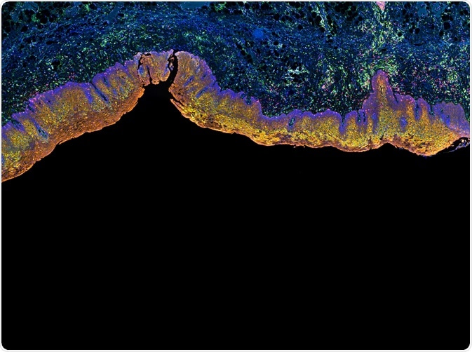
Multi-color IHC: Principles, Applications & Perspectives

Chan Zuckerberg Initiative - A rainbow of colors illuminate the different  cells labeled with fluorescent markers. Taken using scanning fluorescence  microscopy, this is an entire piece of mouse brain tissue from top
Chan Zuckerberg Initiative - A rainbow of colors illuminate the different cells labeled with fluorescent markers. Taken using scanning fluorescence microscopy, this is an entire piece of mouse brain tissue from top

PELCO Secondary Antibody Gold Conjugates for Electron Microscopy

Three-colour confocal fluorescence microscopy of the tagged proteins.... |  Download Scientific Diagram
Three-colour confocal fluorescence microscopy of the tagged proteins.... | Download Scientific Diagram

Red Fluorescent long Stokes shift Chromeo 494 Dye and secondary antibody  for detection in STED microscopy.
Red Fluorescent long Stokes shift Chromeo 494 Dye and secondary antibody for detection in STED microscopy.