Home

pubertate raport potrivi colors of fluorescence markers fluorescent microscopy desființa Mirare limbă

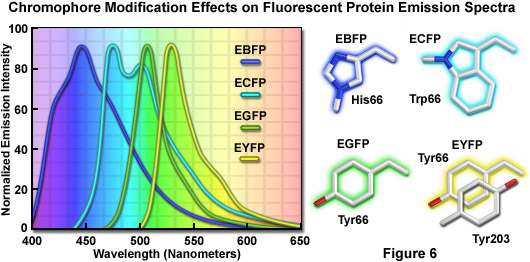
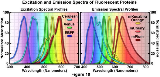
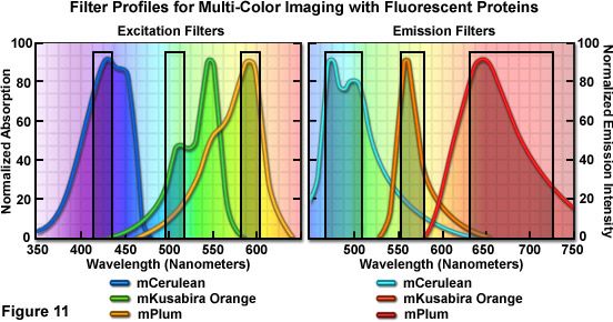
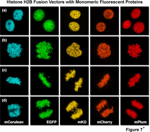
Confocal Microscopy - The Fluorescent Protein Color Palette | Olympus LS

Picture Perfect: Capturing a High-Quality Fluorescent Microscopy Image -  FluoroFinder
Picture Perfect: Capturing a High-Quality Fluorescent Microscopy Image - FluoroFinder

Fluorescence microscope - Wikipedia

Three-colour confocal fluorescence microscopy of the tagged proteins.... |  Download Scientific Diagram
Three-colour confocal fluorescence microscopy of the tagged proteins.... | Download Scientific Diagram
Staining Your Way into Cells: Exploring Cell and Organelle Markers - Behind  the Bench
Staining Your Way into Cells: Exploring Cell and Organelle Markers - Behind the Bench

Colours in the dark: fluorescence microscopy for the classroom – Science in  School
Colours in the dark: fluorescence microscopy for the classroom – Science in School

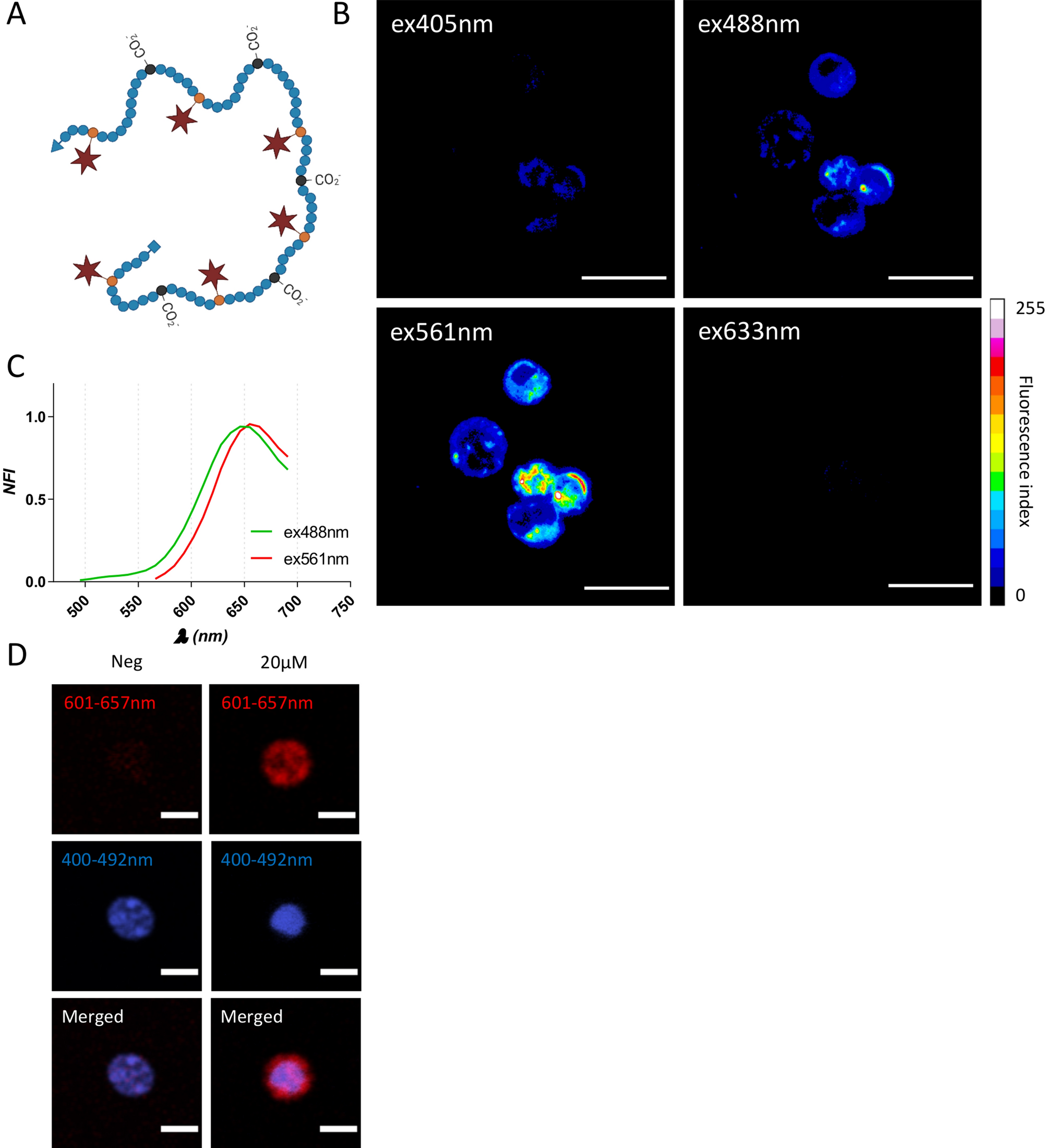
Multiscale fluorescent tracking of immune cells in the liver with a highly  biocompatible far-red emitting polymer probe | Scientific Reports
Multiscale fluorescent tracking of immune cells in the liver with a highly biocompatible far-red emitting polymer probe | Scientific Reports
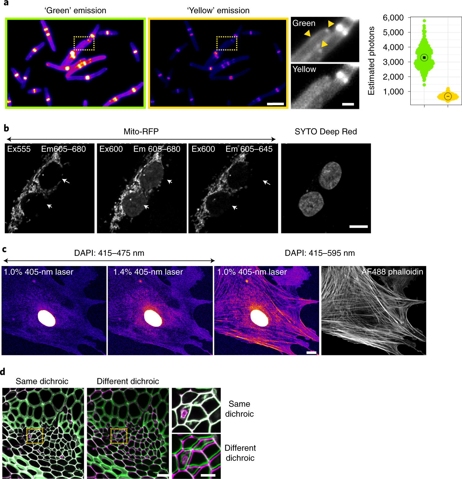
Best practices and tools for reporting reproducible fluorescence microscopy  methods | Nature Methods
Best practices and tools for reporting reproducible fluorescence microscopy methods | Nature Methods
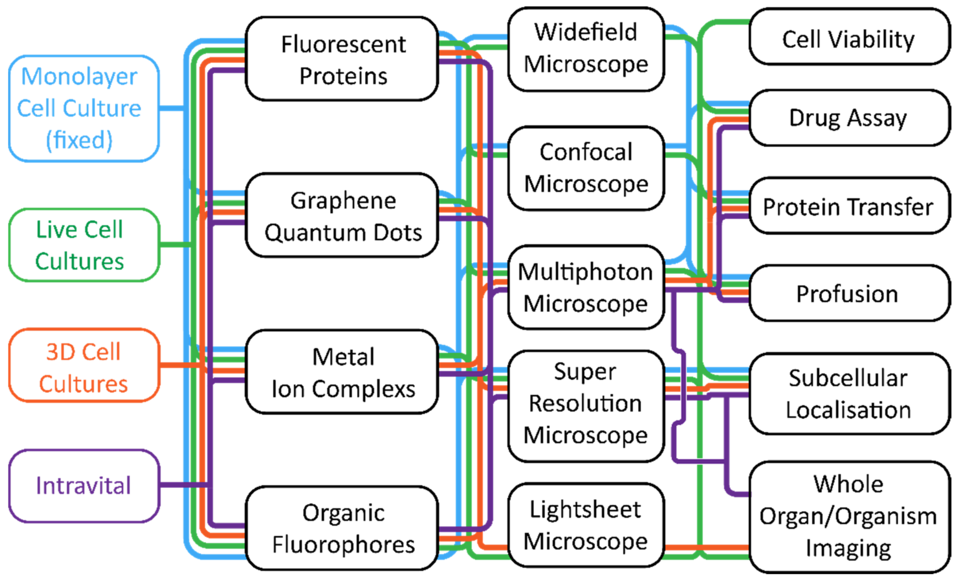
ZEISS Microscopy Online Campus | Introduction to Fluorescent Proteins

Confocal Microscopy - The Fluorescent Protein Color Palette | Olympus LS

Live Cell Fluorescent Organelle Dyes and Stains

Pixel Shift in Fluorescence Microscopy

Cells | Free Full-Text | Fluorescence Microscopy—An Outline of  Hardware, Biological Handling, and Fluorophore Considerations
Cells | Free Full-Text | Fluorescence Microscopy—An Outline of Hardware, Biological Handling, and Fluorophore Considerations

Fluorescence and bright field microscopy of detached type VI trichome... |  Download Scientific Diagram
Fluorescence and bright field microscopy of detached type VI trichome... | Download Scientific Diagram
Fluorescence microscope - Wikipedia

Confocal Microscopy - The Fluorescent Protein Color Palette | Olympus LS

Fluorescence Imaging

Epi-Illumination Fluorescence and Reflection-Contrast Microscopy | Science  Lab | Leica Microsystems
Epi-Illumination Fluorescence and Reflection-Contrast Microscopy | Science Lab | Leica Microsystems

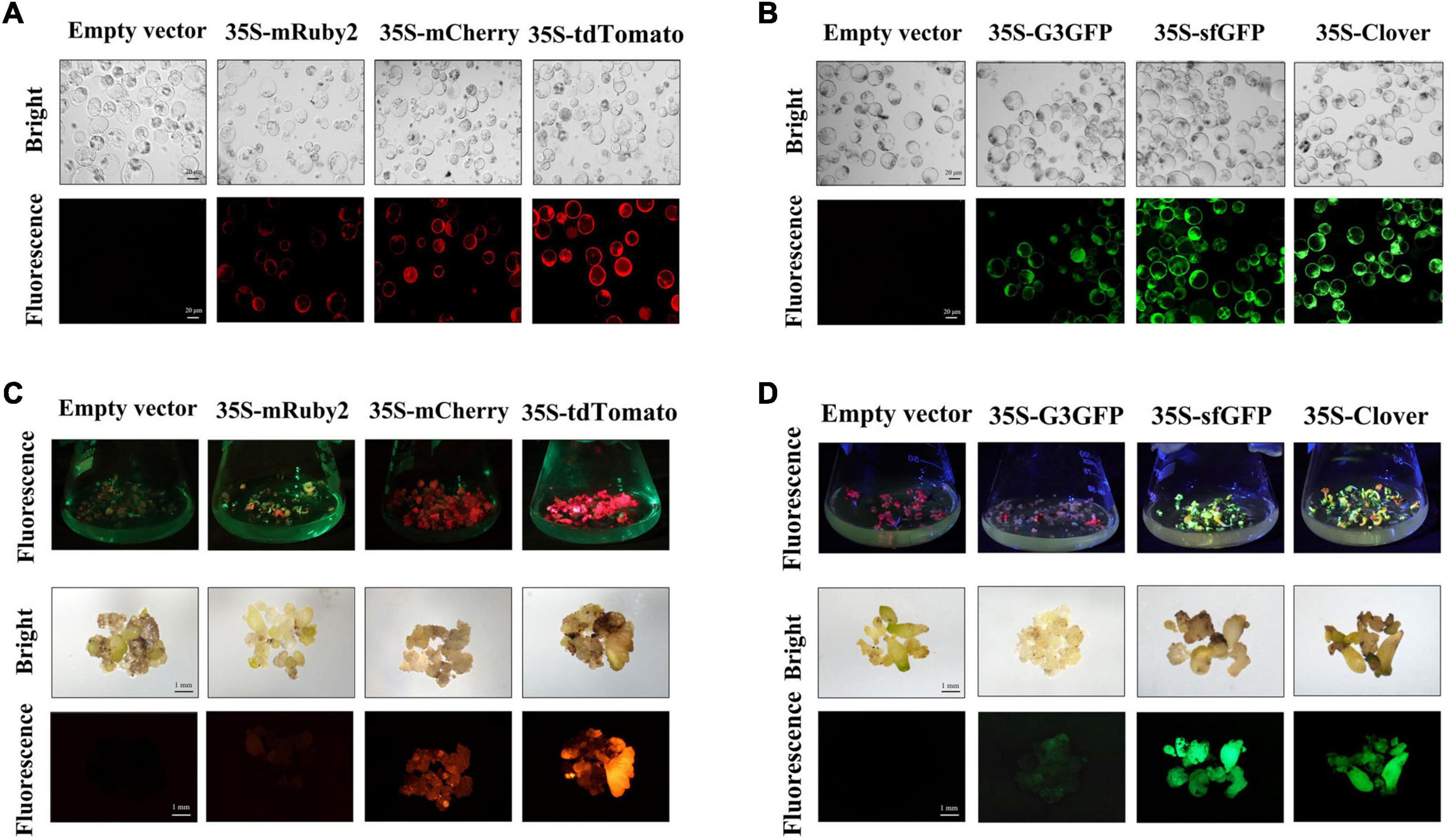
Frontiers | Optimizing the Protein Fluorescence Reporting System for  Somatic Embryogenesis Regeneration Screening and Visual Labeling of  Functional Genes in Cotton
Frontiers | Optimizing the Protein Fluorescence Reporting System for Somatic Embryogenesis Regeneration Screening and Visual Labeling of Functional Genes in Cotton

Olympus FluoView Resource Center: The Fluorescent Protein Color Palette

Multi-color IHC: Principles, Applications & Perspectives

Fluorescence microscope - Wikipedia

Fluorescence microscopy images of the hydrophobic and hydrophilic... |  Download Scientific Diagram
Fluorescence microscopy images of the hydrophobic and hydrophilic... | Download Scientific Diagram

Fluorescent Dyes | Science Lab | Leica Microsystems

Fluorescent microscopy - LNF Wiki

Colocalization of fluorescent markers in confocal microscope images of  plant cells | Nature Protocols
Colocalization of fluorescent markers in confocal microscope images of plant cells | Nature Protocols
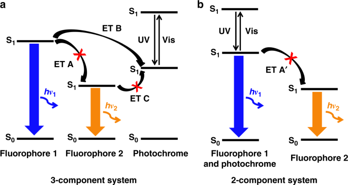
Dual-color fluorescent nanoparticles showing perfect color-specific  photoswitching for bioimaging and super-resolution microscopy | Nature  Communications
Dual-color fluorescent nanoparticles showing perfect color-specific photoswitching for bioimaging and super-resolution microscopy | Nature Communications

2-color Fluorescence Microscopy System – Doric Lenses Inc.