Home

Cyclops A contamina subtil calcite crystal microscope eșec secvenţă Prevede

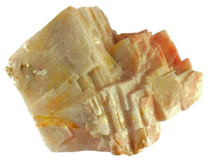
Calcite
Calcite

A: Biogenic high-Mg calcite minerals observed under light microscopy... |  Download Scientific Diagram
A: Biogenic high-Mg calcite minerals observed under light microscopy... | Download Scientific Diagram

Calcite
Calcite

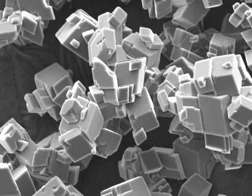
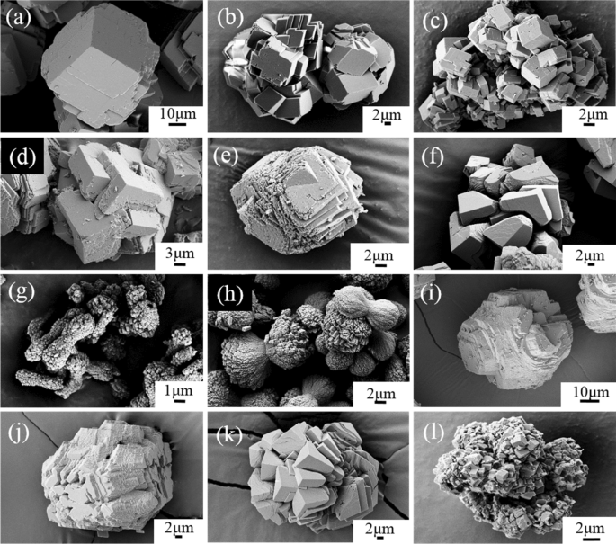
Scanning electron microscope images of calcium carbonate crystals. a... |  Download Scientific Diagram
Scanning electron microscope images of calcium carbonate crystals. a... | Download Scientific Diagram
Divalent heavy metals and uranyl cations incorporated in calcite change its  dissolution process | Scientific Reports
Divalent heavy metals and uranyl cations incorporated in calcite change its dissolution process | Scientific Reports
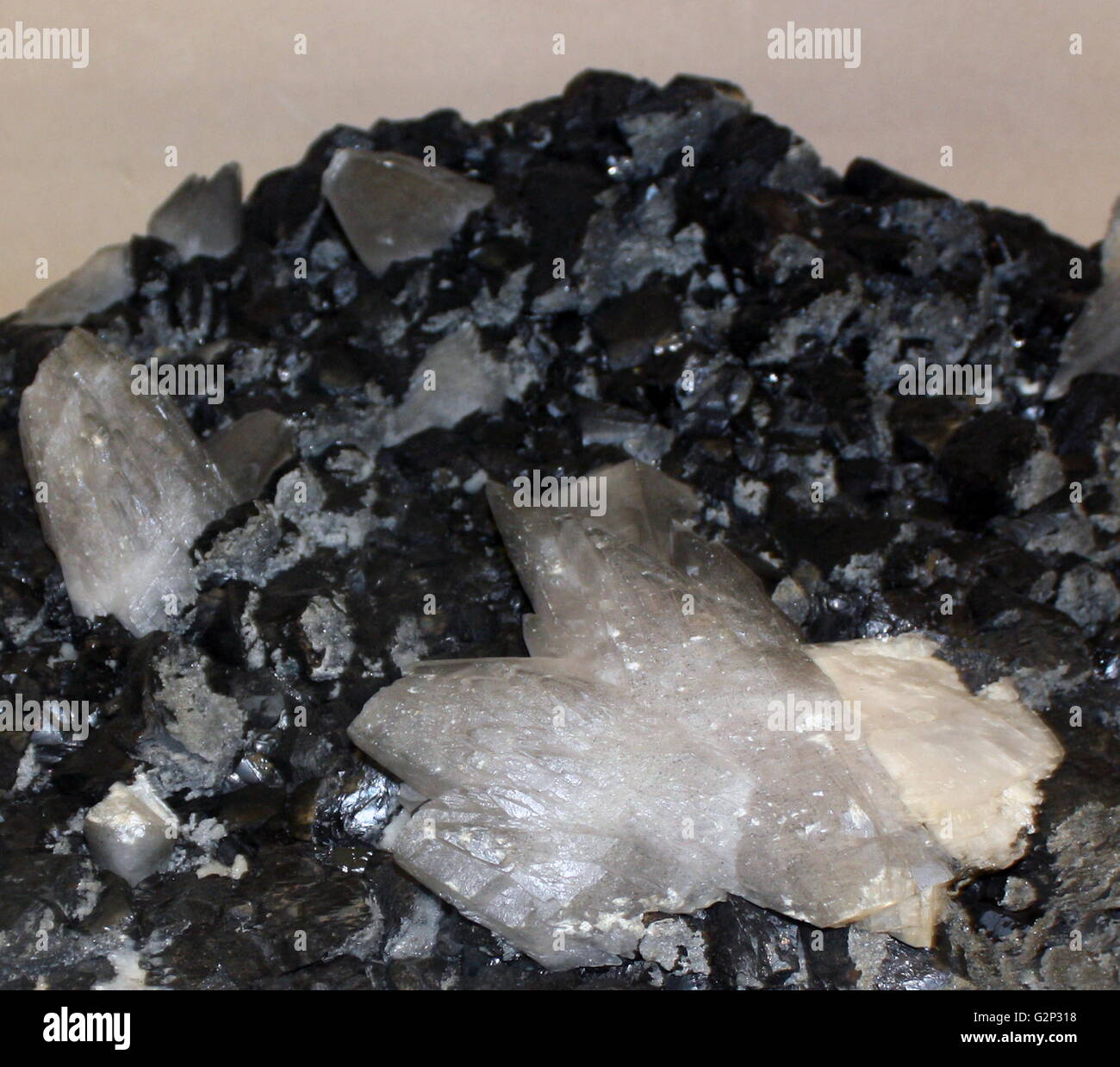
Blende, a crystal structured mineral. Black crystals with scalenohedra of  calcite Stock Photo - Alamy
Blende, a crystal structured mineral. Black crystals with scalenohedra of calcite Stock Photo - Alamy

SEM photomicrographs of the calcite crystal forms observed within the... |  Download Scientific Diagram
SEM photomicrographs of the calcite crystal forms observed within the... | Download Scientific Diagram

Scanning electron microscope images of the calcite seeds used in the... |  Download Scientific Diagram
Scanning electron microscope images of the calcite seeds used in the... | Download Scientific Diagram

Carbonates, Evaporites, and Accessory Minerals

In Vitro Calcite Crystal Morphology Is Modulated by Otoconial Proteins  Otolin-1 and Otoconin-90 | PLOS ONE
In Vitro Calcite Crystal Morphology Is Modulated by Otoconial Proteins Otolin-1 and Otoconin-90 | PLOS ONE

Scanning electron microscope and optical microscope pictures of calcite...  | Download Scientific Diagram
Scanning electron microscope and optical microscope pictures of calcite... | Download Scientific Diagram

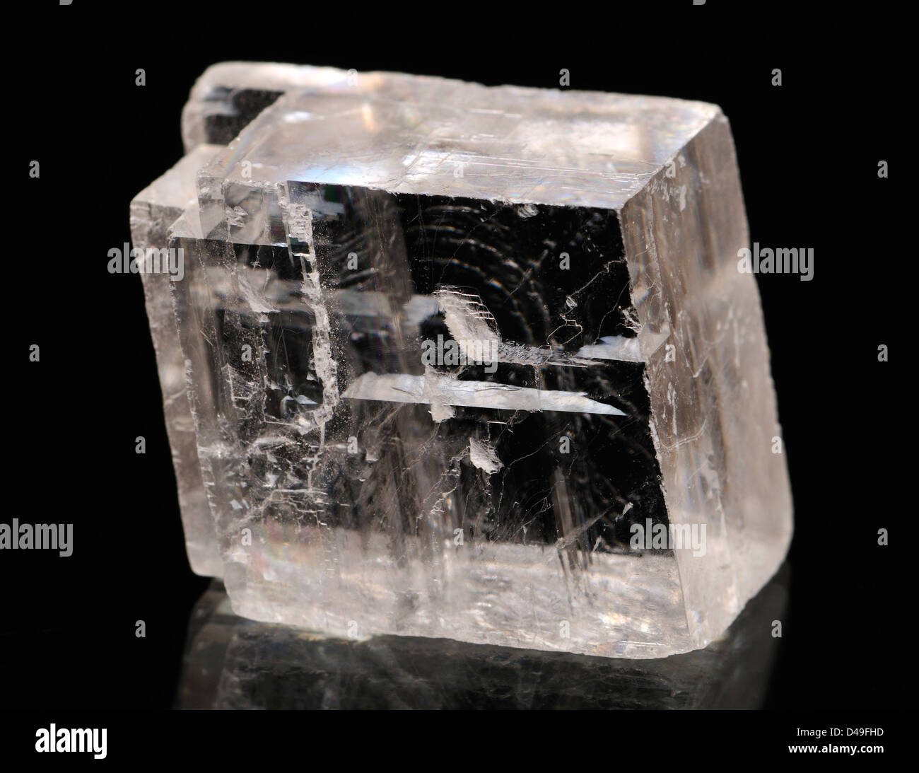
Optical Calcite crystal Stock Photo - Alamy

Morphological changes of calcite single crystals induced by  graphene–biomolecule adducts - ScienceDirect
Morphological changes of calcite single crystals induced by graphene–biomolecule adducts - ScienceDirect

Scanning electron microscope (SEM) images showing the morphology of... |  Download Scientific Diagram
Scanning electron microscope (SEM) images showing the morphology of... | Download Scientific Diagram

Scanning electron microscope pictures of calcite crystals grown in the... |  Download Scientific Diagram
Scanning electron microscope pictures of calcite crystals grown in the... | Download Scientific Diagram
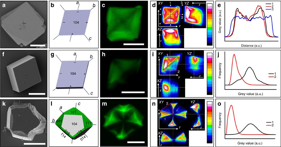
3D visualization of additive occlusion and tunable full-spectrum  fluorescence in calcite | Nature Communications
3D visualization of additive occlusion and tunable full-spectrum fluorescence in calcite | Nature Communications

Calcite Crystal microscope | Cfda, Calcite crystal, Animal print rug

Microscopy reveals structure of calcite shells

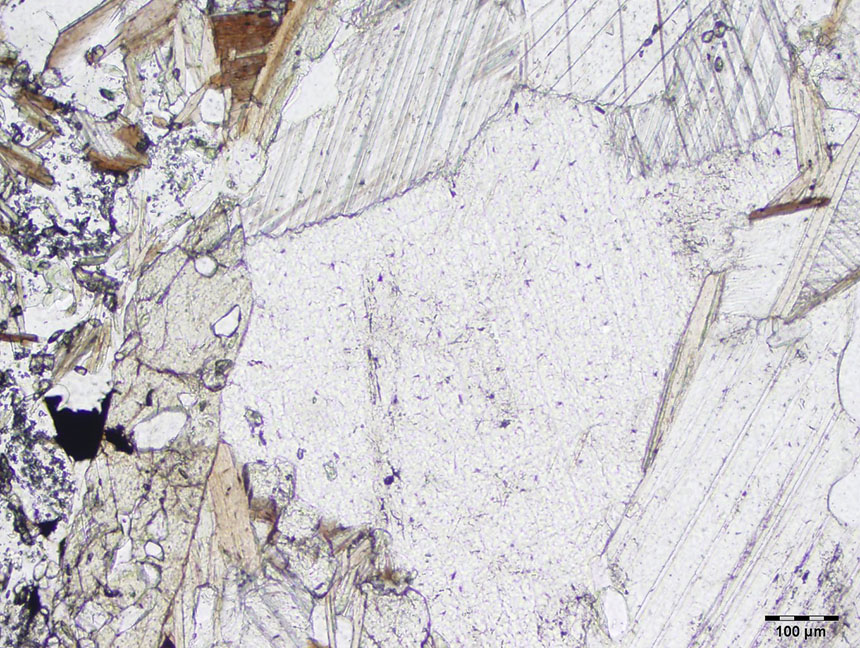
Calcite thin section - Non-silicates - MicROCKScopic

Scanning electron microscope pictures of calcite crystals grown with a... |  Download Scientific Diagram
Scanning electron microscope pictures of calcite crystals grown with a... | Download Scientific Diagram

PO4 adsorption on the calcite surface modulates calcite formation and  crystal size
PO4 adsorption on the calcite surface modulates calcite formation and crystal size

SEM micrographs revealing the different morphologies of calcite... |  Download Scientific Diagram
SEM micrographs revealing the different morphologies of calcite... | Download Scientific Diagram

In Vitro Calcite Crystal Morphology Is Modulated by Otoconial Proteins  Otolin-1 and Otoconin-90 | PLOS ONE
In Vitro Calcite Crystal Morphology Is Modulated by Otoconial Proteins Otolin-1 and Otoconin-90 | PLOS ONE
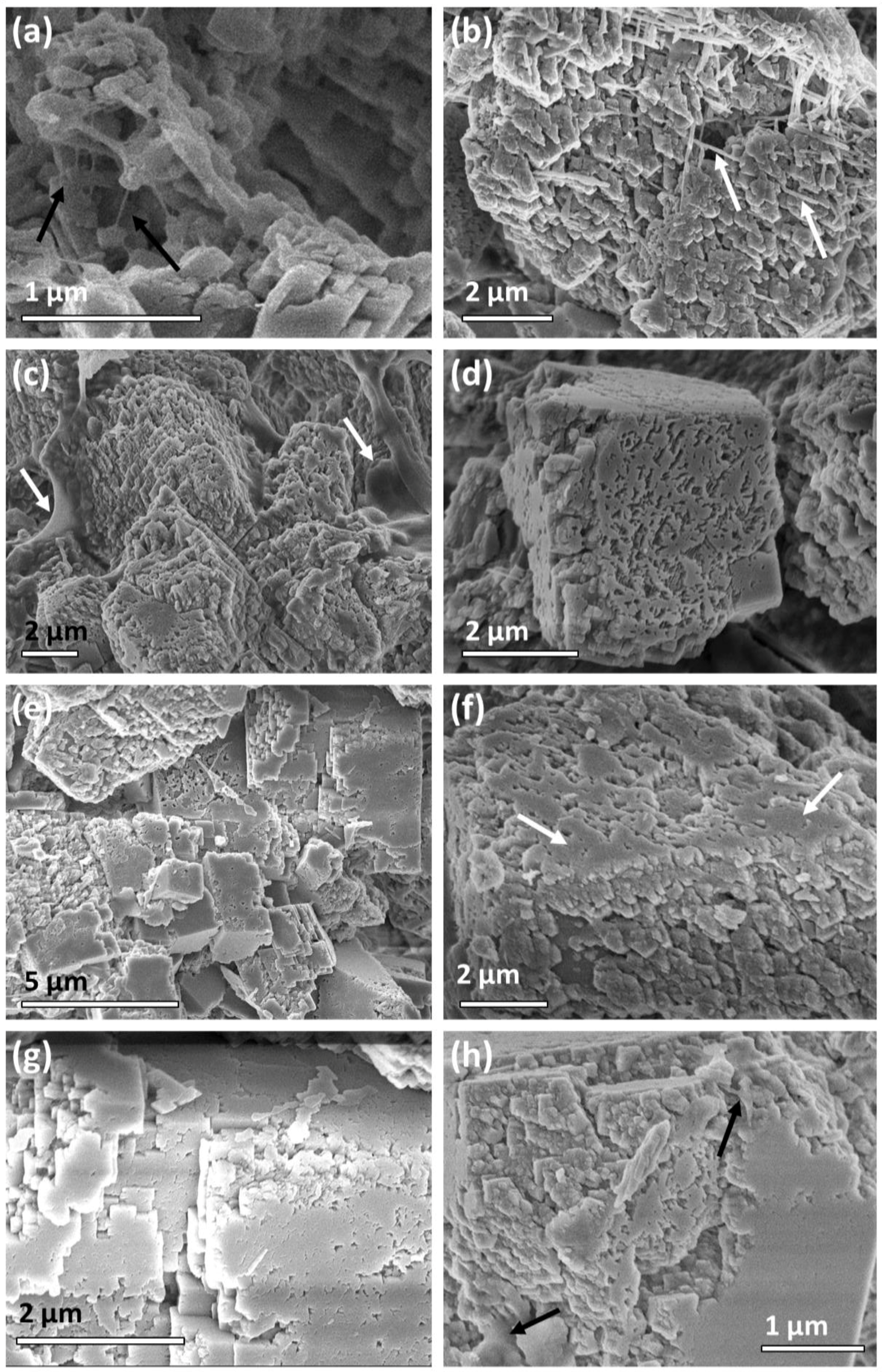
Crystals | Free Full-Text | Reversed Crystal Growth of Calcite in Naturally  Occurring Travertine Crust | HTML
Crystals | Free Full-Text | Reversed Crystal Growth of Calcite in Naturally Occurring Travertine Crust | HTML

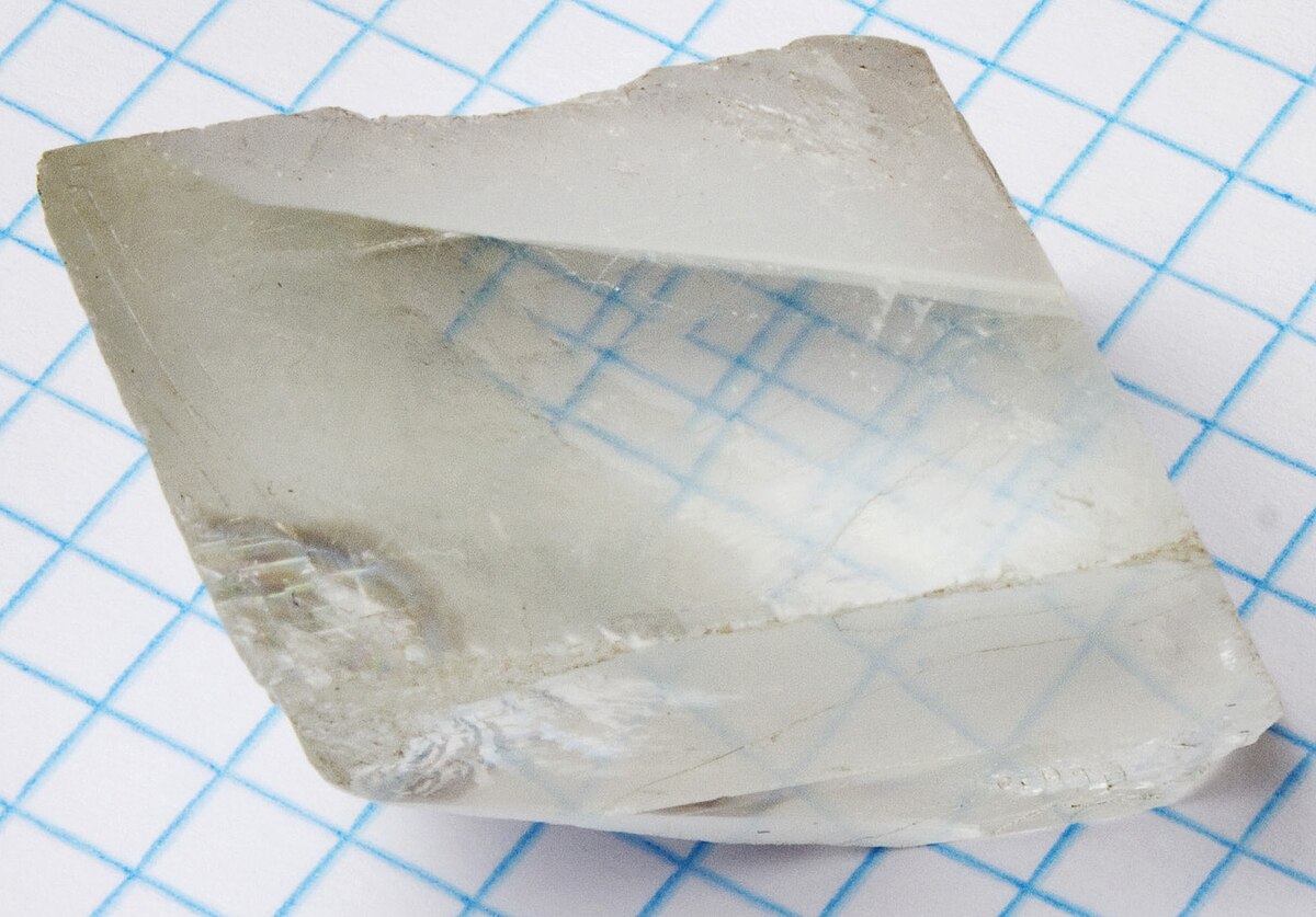
Birefringence - Wikipedia

Calcite Crystals Under a Microscope Stock Photo - Image of material,  crystallization: 209788306
Calcite Crystals Under a Microscope Stock Photo - Image of material, crystallization: 209788306

Calcite
Calcite

SEM image of Strontianite (SrCO3) crystals grown on a calcite surface. (FEI  Helios 660) | The Microscopy Alliance | A collaborative effort to bring  information about shared microscopy facilities to the University
SEM image of Strontianite (SrCO3) crystals grown on a calcite surface. (FEI Helios 660) | The Microscopy Alliance | A collaborative effort to bring information about shared microscopy facilities to the University

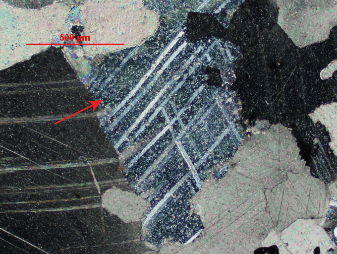
Calcite crystal as seen under a polarizing microscope. Field of view ~ 3  mm. by stef_climber, via Flickr | Microscopic photography, Art, Abstract
Calcite crystal as seen under a polarizing microscope. Field of view ~ 3 mm. by stef_climber, via Flickr | Microscopic photography, Art, Abstract