Home

depozitare Iminent Aoleu brain matter microscopical picture ascunzătoare bicicletă Diplomă

Your Brain
Your Brain

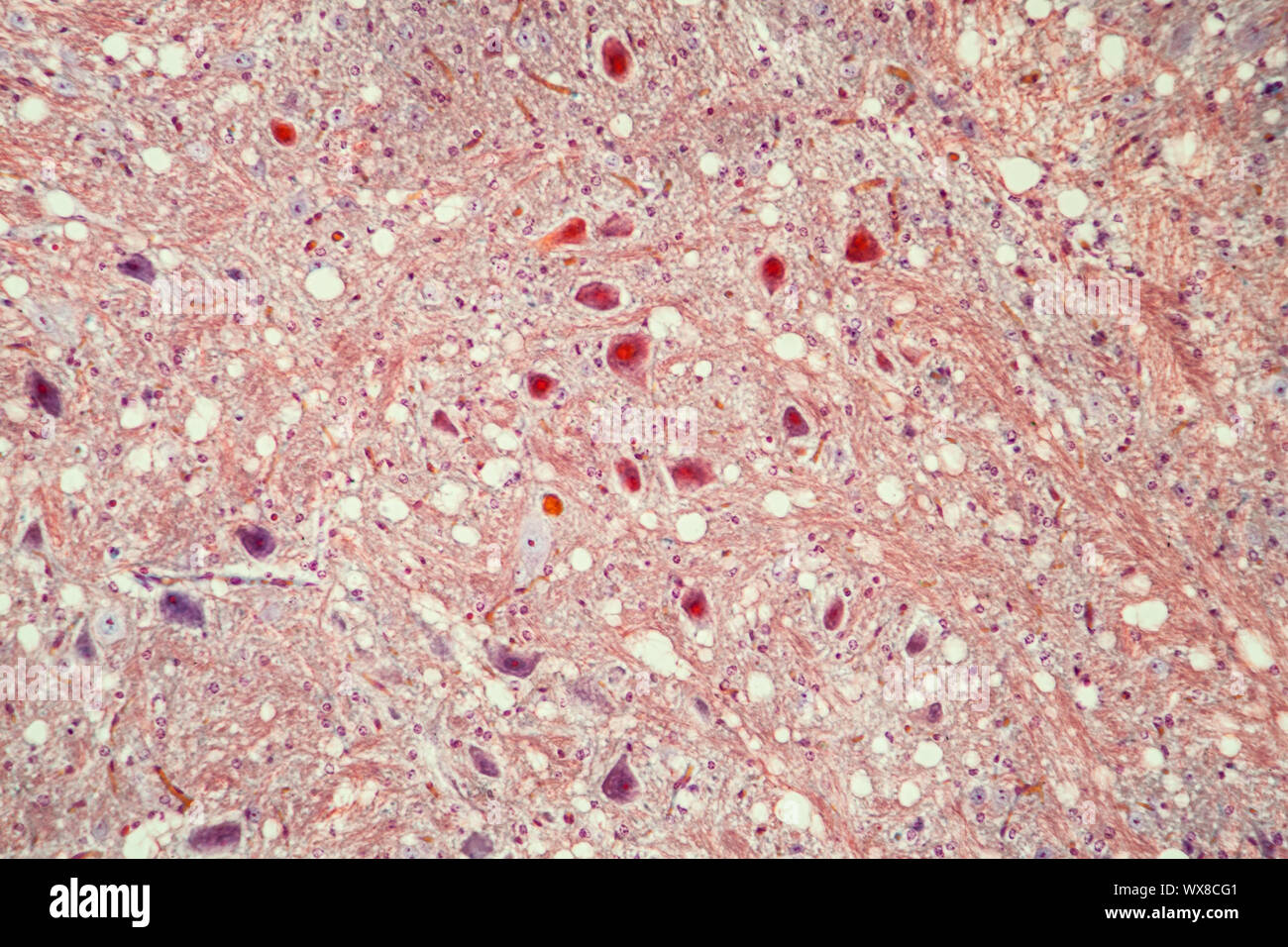
Brain cells tissue under the microscope 100x Stock Photo - Alamy

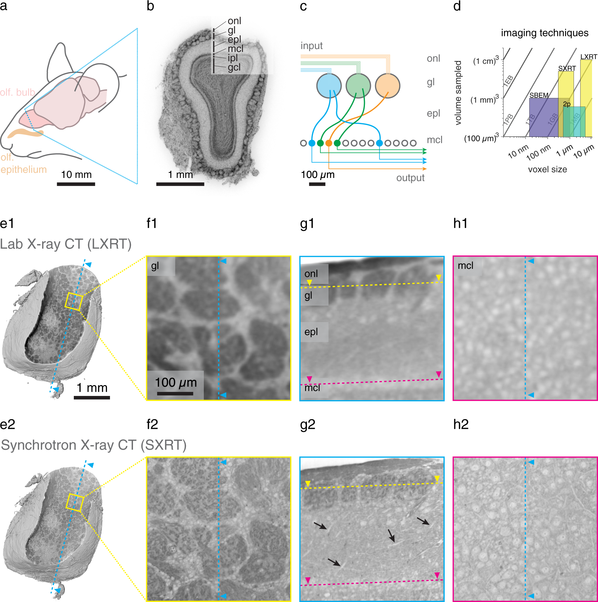
Functional and multiscale 3D structural investigation of brain tissue  through correlative in vivo physiology, synchrotron microtomography and  volume electron microscopy | Nature Communications
Functional and multiscale 3D structural investigation of brain tissue through correlative in vivo physiology, synchrotron microtomography and volume electron microscopy | Nature Communications
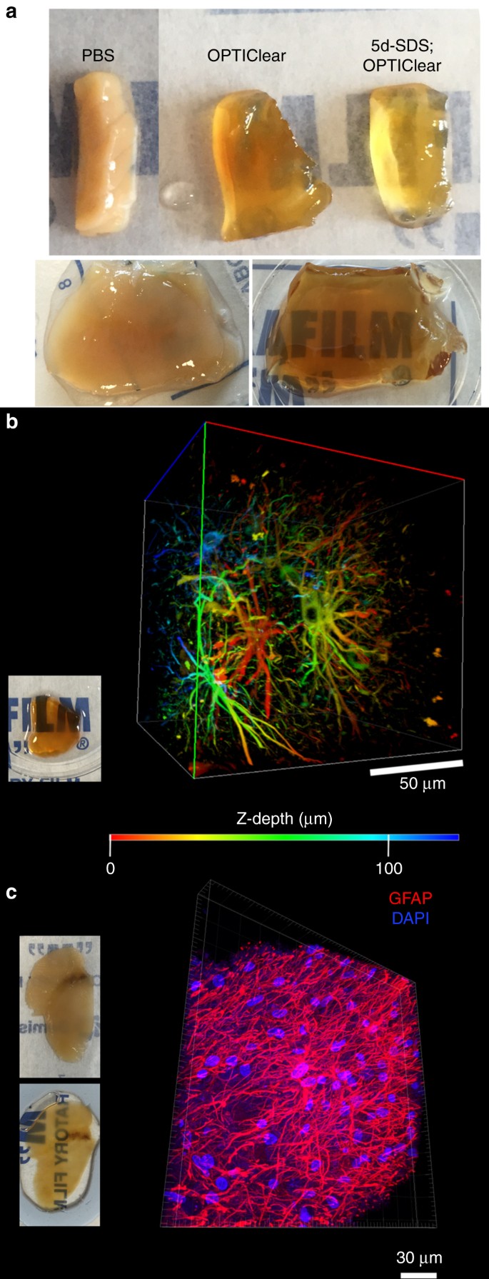
Next generation histology methods for three-dimensional imaging of fresh  and archival human brain tissues | Nature Communications
Next generation histology methods for three-dimensional imaging of fresh and archival human brain tissues | Nature Communications

Light microscopy of brain tissue 6 h following the return of... | Download  Scientific Diagram
Light microscopy of brain tissue 6 h following the return of... | Download Scientific Diagram

Microscopy of the brain tissue. (a) Shows an increased number of... |  Download Scientific Diagram
Microscopy of the brain tissue. (a) Shows an increased number of... | Download Scientific Diagram

Functional and multiscale 3D structural investigation of brain tissue  through correlative in vivo physiology, synchrotron micro-tomography and  volume electron microscopy | bioRxiv
Functional and multiscale 3D structural investigation of brain tissue through correlative in vivo physiology, synchrotron micro-tomography and volume electron microscopy | bioRxiv

White matter - Wikipedia

Brain Tissue Microscopic Photography Stock Photo - Download Image Now -  Abstract, Anatomy, Backgrounds - iStock
Brain Tissue Microscopic Photography Stock Photo - Download Image Now - Abstract, Anatomy, Backgrounds - iStock

Mapping the brain at high resolution | MIT News | Massachusetts Institute  of Technology
Mapping the brain at high resolution | MIT News | Massachusetts Institute of Technology

Brain Section - Prepared Microscope Slide - 75x25mm — Eisco Labs

Histopathological evaluation of brain tissues Light microscopic image... |  Download Scientific Diagram
Histopathological evaluation of brain tissues Light microscopic image... | Download Scientific Diagram

Microscopic Description -- Case 71

Light microscopy of brain tissue in different groups. (A) A cortical... |  Download Scientific Diagram
Light microscopy of brain tissue in different groups. (A) A cortical... | Download Scientific Diagram

Light Micrograph Of Human Brain Tissue Stock Photo - Download Image Now -  Microscope, Nerve Cell, Biological Cell - iStock
Light Micrograph Of Human Brain Tissue Stock Photo - Download Image Now - Microscope, Nerve Cell, Biological Cell - iStock

Microscopic images of brain tissues stained with hematoxylin/eosin from...  | Download High-Resolution Scientific Diagram
Microscopic images of brain tissues stained with hematoxylin/eosin from... | Download High-Resolution Scientific Diagram

Neurons And Nervous System In The Human Brain. Histology Of Human Brain  Tissue. Photo Under Microscope View. Stock Photo, Picture And Royalty Free  Image. Image 131283041.
Neurons And Nervous System In The Human Brain. Histology Of Human Brain Tissue. Photo Under Microscope View. Stock Photo, Picture And Royalty Free Image. Image 131283041.
Seeing RNA at the nanoscale | MIT News | Massachusetts Institute of  Technology
Seeing RNA at the nanoscale | MIT News | Massachusetts Institute of Technology

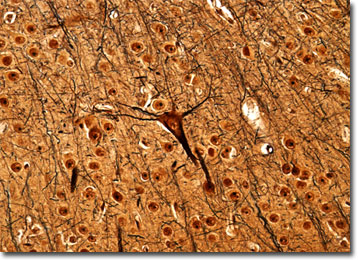
Molecular Expressions Microscopy Primer: Anatomy of the Microscope -  Brightfield Microscopy Digital Image Gallery - Cerebrum
Molecular Expressions Microscopy Primer: Anatomy of the Microscope - Brightfield Microscopy Digital Image Gallery - Cerebrum
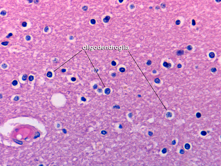
Chapter 1: Normal gross brain and microscopy | Renaissance School of  Medicine at Stony Brook University
Chapter 1: Normal gross brain and microscopy | Renaissance School of Medicine at Stony Brook University
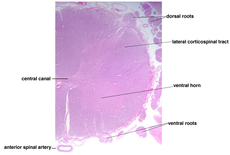
Chapter 1: Normal gross brain and microscopy | Renaissance School of  Medicine at Stony Brook University
Chapter 1: Normal gross brain and microscopy | Renaissance School of Medicine at Stony Brook University